
Loss of Cerebral Volume on MRI in a Child with Severe Iron Deficiency Anaemia: Case Repot and Systematic Review of the literature.
2Community Paedaitrics, Virgincare, UK
Background: A 2 year old girl presented to hospital with severe lethargy, pallor and increased work of breathing on a background of regression of motor and cognitive skills; having previously been able to walk she was now unable to weight bear. The girl was drinking up to 2 litres of cow’s milk daily and eating little solid food.
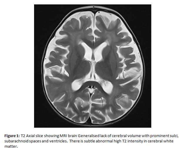
Blood tests revealed a haemoglobin of 11.7g/L (94-130) MCV 63f/L (87-103) and ferritin of 12ug/L (15-150). She was stabilised with a blood transfusion. Echocardiogram showed mild left ventricular dysfunction alongside a pericardial effusion. MRI brain revealed generalised volume loss with prominent sulci, subarachnoid spaces and ventricles (see figure 1). After investigations into potential haematological, metabolic or malabsorptive causes, a diagnosis of severe iron deficiency anaemia was made.
Objective: Iron deficiency anaemia is the most common mineral deficiency of childhood (WHO 2017) and is known to be associated with cognitive impairment and developmental delay (Lozoff 2006). Iron is essential for brain development and iron deficiency has been demonstrated to produce structural brain changes on MRI in animal models (Mudd 2018). We wondered if there were any previous studies finding structural brain changes in children as a result of iron deficiency anaemia.
Methods: Systematic literature review
Results: We found no evidence of any previous studies reporting structural MRI changes. One case series (Munot 2011) reported on 4 children aged 14 months to 4 years with ischemic stroke as a result of IDA.
Conclusion: While it has been shown that iron deficiency produces developmental impairment, it has yet to be elucidated how the deficiency directly affects the brain in children. This case shows a severe case of Iron deficiency anaemia with associated widespread volume loss. Further neuroimaging studies are required to characterize the effect of iron deficiency anaemia on the brain in childhood.
Powered by Eventact EMS