
The Investigation of Mediastinal Opacity in A 10 Years Old Boy Presenting with Asthma Exacerbation
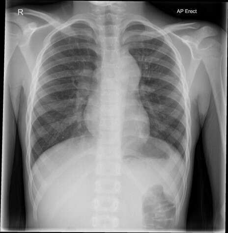
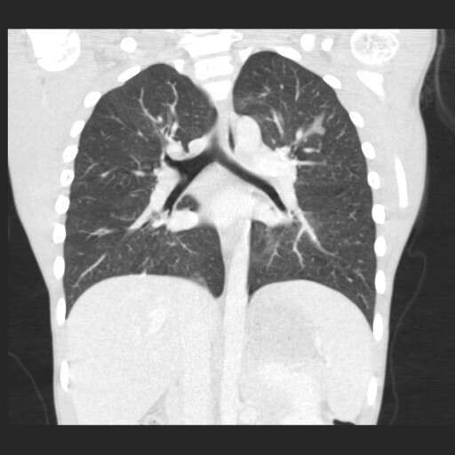
Case: A 10 years old boy, known to have asthma, house dust-mite allergy and unilateral dysplastic kidney, presented to A&E with an exacerbation. His chest radiograph showed bulky mediastinum so was referred to respiratory team. He was treated for atypical infection and echocardiography and abdominal USS were done and were unremarkable. CT chest showed mediastinal lymph node enlargement. The case was discussed with oncology/radiology teams and agreed on treating as atypical infection. Serial chest radiographs revealed gradual improvement, then was lost for follow up for 10 months. When he was seen in clinic, a chest X-ray showed that the hilar prominence is still there, but was asymptomatic, and later on, in 2 months the changes were persistent, so he went on to have a thoracic MRI that revealed a mediastinal mass likely lymphadenopathy with a splenic lesion and an abnormal marrow signal along the spine. Splenic and bone marrow biopsies both showed no malignant cells. A PET scan revealed activity consistent with lymphoma that was later confirmed on axillary lymph node biopsy and the boy was subsequently started on chemotherapy.
Conclusions and learning points:
This case shows that it can be difficult to interpret mediastinal opacities on plain radiographs and in this case the CT scan has not changed the management significantly.
PET scan helped guide further investigations.
Biopsy of an appropriate site is the gold standard (as two biopsies from spleen and bone marrow were negative), and in this particular case, would have saved much effort and time.
Powered by Eventact EMS