
Mediastinal Mass Presenting as Suspected Dysfunctional Breathing in A 14 Years Old Girl
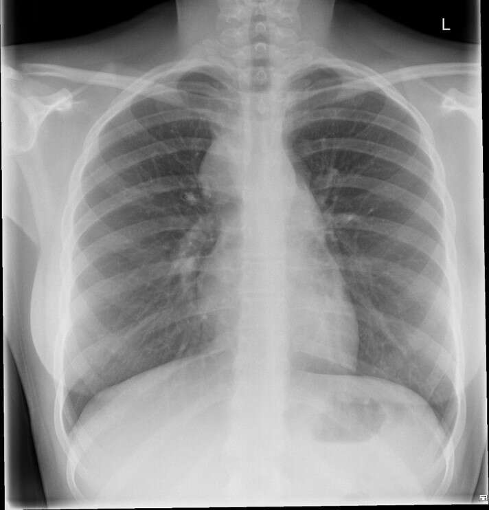
A 14 years old girl, known to have autism and eczema, was referred to general paediatrics by her GP with history of right lower chest pain for 2 years. Pain has worsened recently, became more central, was aggravated by exercise and with no relieving factors. Her clinical examination was normal. Chest X-ray was unremarkable, the pain was thought to be of musculoskeletal origin. A trial of salbutamol inhaler and antacid did not help her symptoms and she was referred to physiotherapy. She was assessed by the physiotherapist who referred her to the respiratory clinic to rule out exercise induced laryngeal obstruction. She was seen in clinic 2 months later, where the chest radiograph was repeated and revealed mediastinal enlargement. Her echocardiography and spirometry were normal. She went on to have a CT chest that confirmed mediastinal lymphadenopathy. A thoracoscopic biopsy confirmed the diagnosis of Hodgkin’s lymphoma. She completed the treatment protocol and subsequent work up showed no residual disease; she is currently asymptomatic under regular follow up.
Conclusions and learning points
This case presented with shortness of breath on exertion which was thought to be due to dysfunctional breathing, which is an unusual presentation of a mediastinal mass.
A normal radiograph at the first presentation should not cause hesitation in repeating it especially with persistent symptoms.
Tissue biopsy is the gold standard investigation in such cases.
Powered by Eventact EMS