Improvement of Renal Function in Transplanted Kidney Patient after Succesful Stenting of Severe Iliac Stenosis, Revealed through Subtle Doppler Changes

Lilian Atlan Ethan Yussim
Radiology, Tel Aviv Sourasky Medical Center, Affiliated to the Sackler School of Medicine, Tel Aviv University, Israel

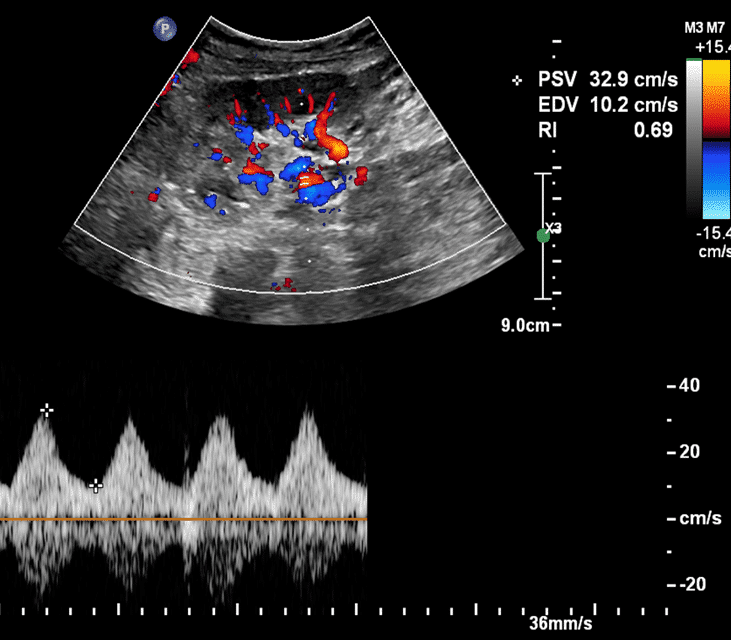
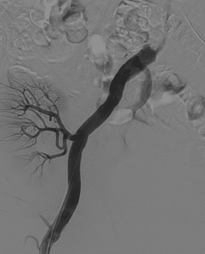
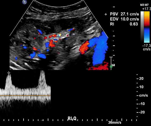
A 74 year old patient with a history of PCKD and kidney transplantation 15 years prior, presented with progressive kidney failure. The ultrasound showed normal size,echogenicity as well as RI values. There was a light delay in the systolic acceleration pattern of the Doppler signal in the proximal kidney arteries. Through angiographic assessment, a severe stenosis was found in the proximal iliac artery, which was treated by stenting. The patient’s Creatinine values improved after the intervention.

Even slight changes in the ultrasound Doppler pattern should raise the suspicion for hemodynamically significant changes in supplying arteries and necessitate further multidisciplinary investigation.

During vascular assessment of the kidney transplant, the Resistive Index ( RI) values were normal in all areas of the transplanted kidney. RI Indexes were within the normal range, between 0.66 to 0.69. No ‘Parvus Tardus’ pattern was present.

Severe stenosis in the ipsilateral common iliac artery

Ballon angioplasty and stent implantation

After the procedure,  full patency of the previously stenotic lesion, as well as good perfusion of the renal arteries up to its distal small branches within the transplant

Follow-Up ultrasound POD1, shows immediate and complete normalization of the Doppler pattern,

Lilian Atlan
Lilian Atlan


Powered by Eventact EMS