Purpose: The aim of this study was to non-invasively assess the relationship between cardiac pH and lactate dehydrogenase (LDH) and pyruvate dehydrogenase (PDH) activities using MRS.
Materials and methods: An isolated perfused mouse heart system was used and the LDH and PDH activities were monitored in real-time by following the production of hyperpolarized [1-13C]lactate and [13C]bicarbonate, respectively, following the administration of hyperpolarized [1-13C]pyruvate to the heart. Cardiac pH and ATP level were determined using 31P MRS.
Retrograde perfusion of the heart, from 5 mice (male, ICR mice, 39-50 g), was performed with modified Krebs-Henseleit buffer in a 10 mm NMR tube, inside a 5.8 T NMR spectrometer. Spectrally selective RF pulses were used to determine the rate of newly formed [1-13C]lactate and [13C]bicarbonate while providing very small excitation of the substrate ([1-13C]pyruvate). Each isolated heart was subjected to 2-3 injections (total number of injections = 11, in 5 hearts). Before each hyperpolarized injection a 31P spectrum was acquired. pH was calculated based on chemical-shift difference of the inorganic phosphate signal (Pi) relative to that of phosphocreatine (PCr).
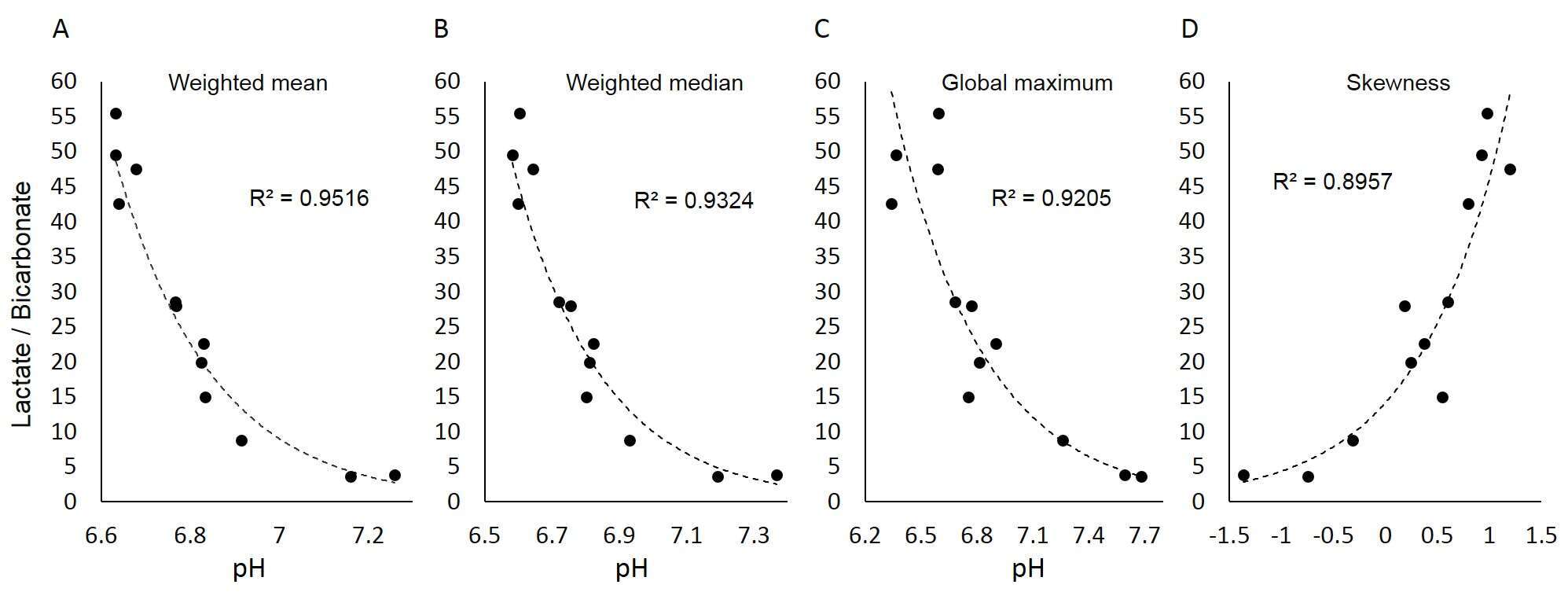
Results: In the retrograde perfused heart model the tissue viability is inadvertently degraded over the course of several hours and the tissue pH is reduced (acidifies). In agreement, a reduction in PCr and all three ATP peaks is observed with time (Figure 1A, bottom to top), while the Pi peak increases and shifts to a lower field with time, indicating tissue viability deterioration (Figure 1A). However, the Pi signal shows multiple components, indicating non-homogenous pH distribution in the cardiac tissue. To describe this heterogeneous pH distribution, we used the weighted mean and its S.D., weighted median, global maximum, skewness, kurtosis and entropy of the pH curve that results from the complex Pi signal. The results indicate that LDH/PDH enzymatic activity ratio is strongly correlated in an exponential manner to the pH of the cardiac tissue. The dependence of the LDH/PDH activity ratio on the mean, median, global maximum, and the skewness of the pH distribution is shown in Figure 2.
Conclusions: We show a correlation between LDH/PDH activities ratio and global aspects of tissue pH in the isolated mouse heart. This finding may offer a new approach for a non-invasive assessment of cardiac pH changes. Further investigation is needed to validate this approach in vivo and understand the mechanistic origin of this correlation.
Figure 1:

Figure 2: