Purpose: Contrast enhanced voiding urosonography (CEVUS) is a sensitive and radiation-free imaging technique for diagnosis and grading of vesico-ureteral reflux (VUR), and for assessment of urethral anomalies. It has become a widely accepted tool in many centers in Europe and United states, as an alternative modality for fluoroscopic voiding cystourography (VCUG). The aim of this study is to describe our initial local experience using CEVUS on detection of VUR and evaluation of the urinary system.
Materials & methods: Retrospective review of the first 53 pediatric CEVUS studies that were performed in Shaare Zedek Medical Center and in Tel-Aviv Sourasky Medical Center, from December 2018 to June 2019.
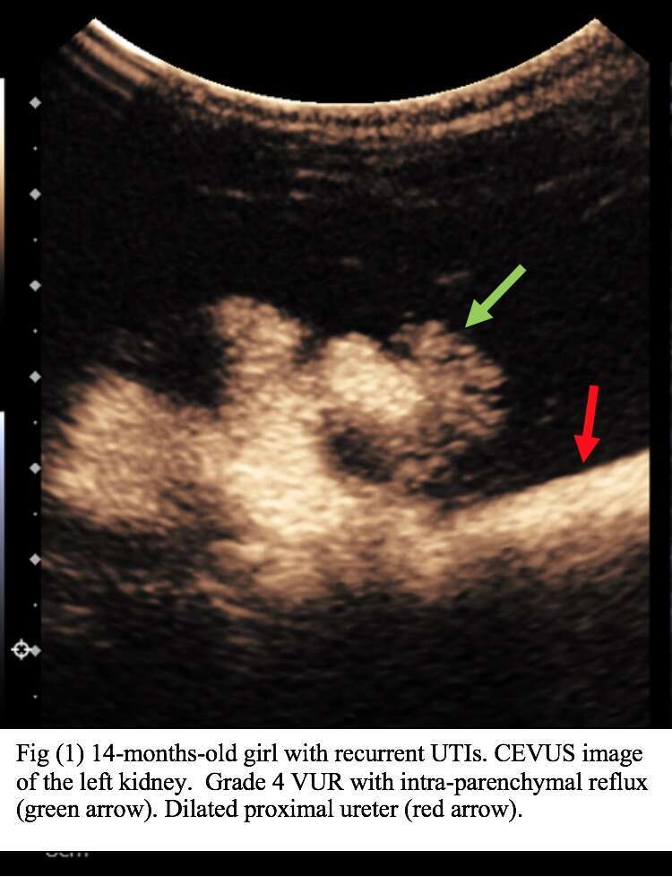
Results: 53 patients (105 pelviureteric units) underwent CEVUS in both medical centers during the study period. Average age was 13.1 months±44.7 (range 2 days-17.7 years). No reflux was found in 21/53 patients (37.7%). Reflux to a single kidney was found in 18 patients (35.9%) (Fig.1), and bilateral reflux was found in 14 patients (26.4%). In 15 patients with reflux, intra-parenchymal reflux was detected (32.6%). The ureters were visualized in 36 of pelviureteric units with reflux (78.3%). The urethra was demonstrated in 40 patients (75.5%). Urethral pathologies were seen in 4 girls (spinning top urethra). Study duration ranged from 6-24 min (average 11.5±3.9). Complications included pyelonephritis in a single patient who did not follow the prophylactic antibiotic protocol. No other complications were noted.
25 patients underwent sedation: 14 had deep sedation by anesthesiologist, and 11 patients had lighter sedation with nasal Midazolam or, in some cases, medical clowns joined the team for anxiolytic purposes.
Conclusions: CEVUS is a simple method for assessment of reflux and other related structural pathologies of the urinary tract. In our experience, the transition from VCUG to CEVUS is intuitive and requires only a short training. The technique has several advantages over VCUG: (1) It is radiation free (2) Our impression is that study duration is shorter compared to VCUG, even in older children. As a result, duration of sedation (when required) is also decreased. Moreover, shorter study time enables examining more patients per day, thus increasing study availability to the patients.