49 y\o women presented with abdominal pain and weight loss for 3 month, accompanied by mild thrombocytopenia and abnormal liver test.
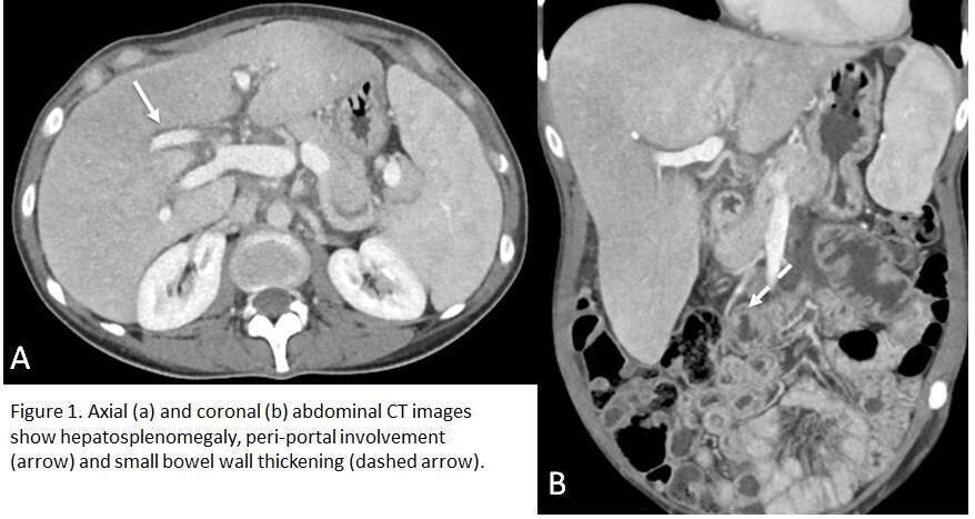
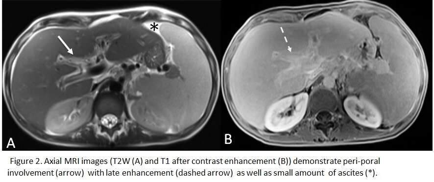
Abdominal CT and MRI findings are shown on figures 1-3.
The working diagnosis included hepatic veno-occlusive disease, lupus and iron overload.
The liver biopsy demonstrated deposition of amyloid, compatible with hepatic amyloidosis.
Teaching points:
- Gastrointestinal system is commonly involved.
- Heterogeneous appearance of the liver, peri-portal involvement, diffuse low signal intensity of spleen on T2-weighted MRI, and thickened bowel wall are suggestive of the diagnosis.
- Biopsy is nearly always required for proper diagnosis.