
Practical Clinical Experience with Patent Ductus Arteriosus Closure Supported by Mixed Reality Interactive Visualization of Cardiovascular Anatomy
Echocardiographic three-dimensional (3D) datasets acquired from transesophageal window are commonly used as procedural aid during percutaneous cardiac interventions. We present initial experience with patent ductus arteriosus (PDA) closure performed with innovative mixed reality display integrated with procedural workflow to improve 3D perception and navigation in 3DTEE datasets.
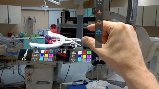
We performed 2 percutaneous occlusions of PDA in adults with aid of intraprocedural mixed-reality display of segmented CTA (computed tomography angiography) data using a voice- and gesture controlled head-mounted display. A dedicated software pathway was used for files conversion, real-time Wi-Fi streaming of 3D rendering from PC to device and manipulation of spatial data during the procedures.
Previously recorded CT angiogram of aorta and ductus was manually segmented and uploaded into 3D DICOM workstation (CarnaLife® Holo, MedApp, Kraków, Poland). 3D holograms were successfully displayed during the procedure by head-mounted display allowing touchless control and image sharing within cath-lab. Wiring of PDA aortic orifice was assisted by 3D hologram controlled by the imaging specialist and shared by the operator. Thus, mixed reality display using the custom software was successfully executed with segmented data presented as a semitransparent cubic hologram positioned in a convenient part of visual field allowing real-world action and with touchless control by medical team. Operator appreciated the use of mixed reality hologram recreating spatial relationships as practical aid to establish anatomical relationships and facilitate entry into ductus orifice. Procedures were successfully completed using arteriovenous guidewire loop to implant Amplatzer Duct II Occluders.
Powered by Eventact EMS