
Nanolayer-controlled faceting of nano- to yocto- liter liquid droplets
2Russell Berrie Nanotechnology Institute (RBNI) and Department of Biotechnology and Food Engineering, Technion – Israel Institute of Technology, Haifa, Israel
Contrary to everyday experience, where all liquid droplets assume rounded, near-spherical shapes, the temperature-tuning of liquid droplets to faceted polyhedral shapes and to spontaneous splitting has been recently demonstrated in oil-in-water emulsions[1]. However, the elucidation of the mechanism driving these surprising effects, as well as their many potential applications, ranging from faceted nanoparticle synthesis, through new industrial emulsification routes, to controlled-release drug delivery within the human body, have been severely hampered by the micron-scale resolution of the light microscopy employed to date in all in situ studies[1-2]. Thus, the thickness of the interfacially frozen crystalline monolayer, suggested to drive these effects, could not be directly measured, and the low limit on the droplet size still showing these effects remained unknown.
We employed a combination of super-resolution stimulated emission depletion microscopy, cryogenic transmission and freeze-fracture electron microscopy, to study these effects well into the nanometer length scale, employing hexadecane oil droplets, stabilized in water by a common cationic surfactant, octadecyltrimethylammonium bromide. We demonstrate the occurrence of the faceting transition in droplets spanning an incredible 12 decades in volume from nanoliters to yoctoliters[3]. We directly visualize the interfacially-frozen, few nanometer thick, crystalline monolayer[3] which drives these effects[1] (Fig. 1). The bulk of the droplets is structureless, ruling out the formation of a surface-adjacent 300 nm - thick crystalline structure, hypothesized in some earlier studies[2] to drive the faceting of the droplets. Finally, our measurements allow placing an upper-limit estimate on the two-dimensional Young modulus of the interfacial nanometer-thick surface crystal in the smallest droplets, providing insights into the virtually unexplored domain of nanoelasticity.
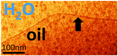
Figure 1: CryoTEM image of the edge of a vitrified faceted droplet. The interfacially-frozen layer, only a few nm thick, is marked by an arrow. No internal structure is visible.
[1] S. Guttman et al., Proc. Natl. Acad. Sci. USA 113 (2016) 493-496.
[2] N. Denkov et al., Nature 528 (2015) 392-396.
[3] S. Guttman et al., Nano Lett. 19 (2019) 3161-3168.
Powered by Eventact EMS