Retrocecal Appendicitis on POCUS: A Case Series
2Pediatric Emergency Medicine, Kaplan Medical Center, ישראל
Pediatric emergency physicians are able to identify appendicitis on point of care ultrasound. An appendix in an unusual location can be challenging to locate, and retrocecal location of the appendix is a risk factor for perforation. We present the first case series of five children diagnosed with retrocecal appendicitis on POCUS in the pediatric emergency department. All had findings consistent with appendicitis on POCUS in the right upper quadrant of the abdomen. Two children were perforated on presentation and one perforated in the operating room. However, in four of the children the white blood cell count was only mildly elevated, in two children the C-Reactive Protein (CRP) was only mildly elevated and in one child the CRP was zero. In two of the children, no appendicitis was found on radiology-performed ultrasound.
POCUS evaluation not only strengthens the PED as the ‘medical home’ for its patients, but expedites diagnosis in children more likely to present late and with perforation, and may decrease radiation exposure from CT. Establishing a POCUS protocol that incorporates both right upper and right lower quadrant views to the examination will decrease the incidence of missed retrocecal appendicitis and may reveal signs of complicated appendicitis.
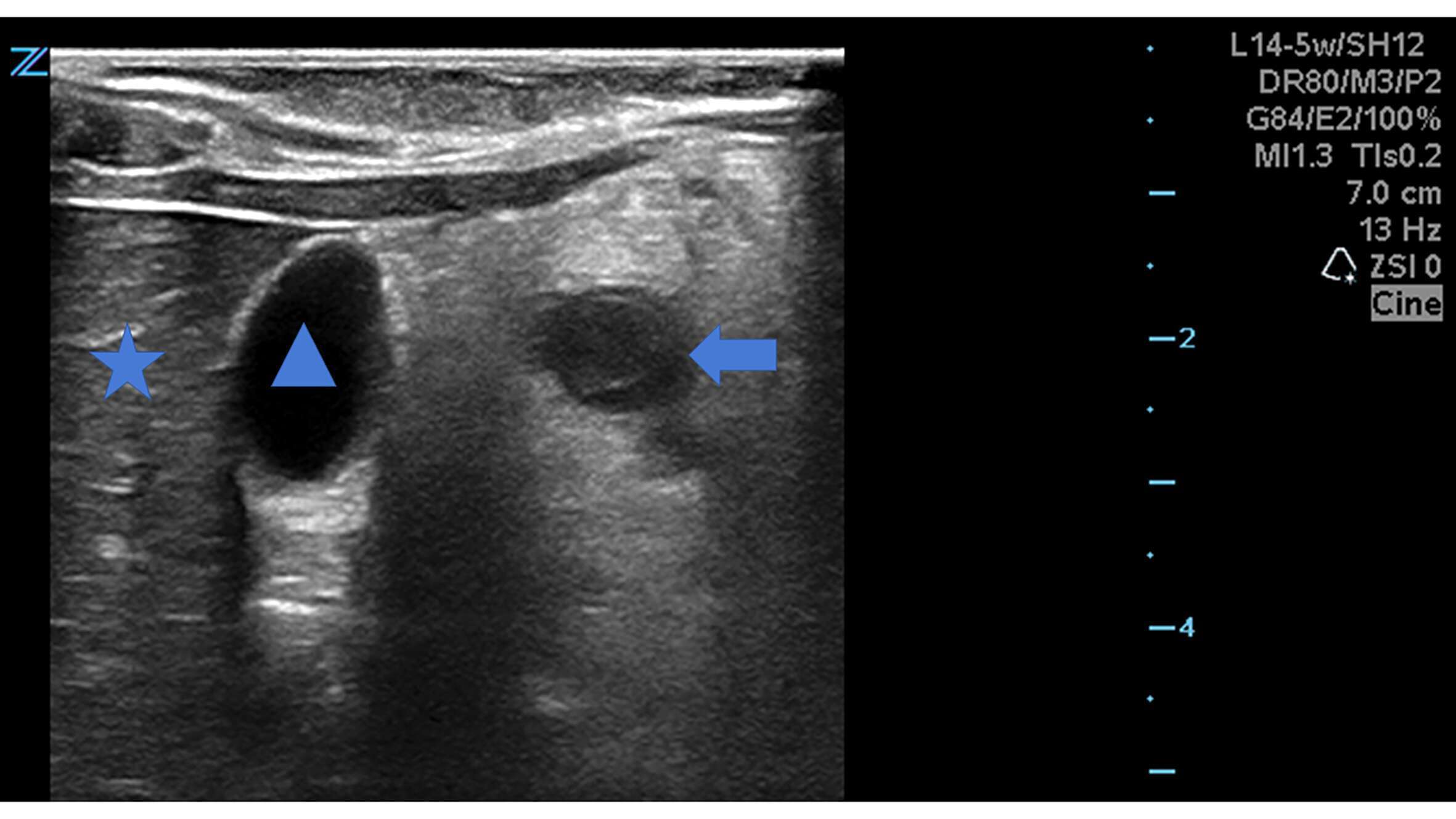
Figure 1:

Legends:
Figure 1: The star sits within the liver, and the triangle within with gallbladder, on a transverse view of the right upper quadrant. The arrow indicates the appendix surrounded by a small amount of hypoechogenic free fluid and echogenic fat.
Similar figures are available for all children in the series.
Powered by Eventact EMS