
First In-Human Implantation of a Wireless Left-Atrial Pressure Sensing System - the VECTOR-HF study
2Careggi, University Hospital, Italy
3Hospital Hamburg-Eppendorf, Hamburg, Germany
4Vectorious Medical Technologies, Tel-Aviv, Israel
5Padeh-Poriya, Medical Center, Israel
6Ohio State University, Columbus OH, USA
7Agostino Gemelli University Policlinic, Rome, Italy
8QEHB, Birmingham, UK
9Vivantes Am-Urban Hospital, Berlin, Germany
10Hammersmith Hospital – Imperial College Healthcare, London, UK
11CardioVascular Center Frankfurt, Frankfurt am Main, Germany
Introduction: Non-invasive methods to assess the risk of heart failure have little specificity and sensitivity for impending exacerbations and subsequent rehospitalizations. So far, only right-sided invasive pressure sensors have been approved for clinical use. Recently, a second-generation monitoring system which assess left-sided pressures, the V-LAP™, has been developed. We hereby describe the first-in-human study.
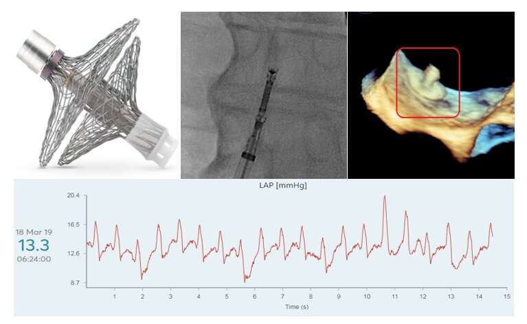
Methods and Results: The V-LAP™ wireless sensor is implanted in a transfemoral, trans-septal fashion, under angiographic and echocardiographic guidance (panels A-C). It resides in the left atrium, where is measures pressure, using digital processing capabilities. The system includes an external home-based unit, which powers the implant by electric inductive coupling, activating it, thus collecting data via radio frequency communication. The remote power induction enables the implant’s battery-less configuration and small size.
Thus far, there have been nine successful implantations of the V-LAP™, performed in NYHA Class III patients (mean age 67.0±8.6, 8/9 male, mean LV ejection fraction – 31.0±5.9%, 6/9 ischemic cardiomyopathy 66.67). All were admitted repeatedly for exacerbations of HF, and demonstrated elevated NT-ProBNP levels. There were no complications. Following implantation of the device, patients were able to perform pressure measurements at home, transmitting data to a secure cloud-centered database. The information gathered includes daily trends and high-resolution waveforms of intracardiac pressures (panel D). This enables assessment of personal response to medications, as well as important clinical events such as atrial arrhythmias and diastolic dysfunction.
Conclusions: We have described the first experience with a digital left-atrial pressure sensor. The ability of the system to assess daily ambulatory hemodynamic information will be demonstrated in the V-LAP monitoring systEm for patients with Chronic sysTOlic and diastolic congestive heart failuRe first-in-human (VECTOR-HF) study.
Panel A The V-LAP™ wireless implant.
Panel B Fluoroscopic images of the implant and delivery system before deployment.
Panel C Transesophageal view of the implant following attachment to the inter-atrial septum.
Panel D High resolution left atrial pressure measurements captured remotely.
Powered by Eventact EMS