
Exuberant Localized Linear Scleroderma and Borrelia Infection: Case Report
2Department of Dermatology, Centro Hospitalar e Universitário de Coimbra, Portugal
Background: Localized linear scleroderma (LLS) is the predominant form of childhood scleroderma, but the initial inflammatory skin lesions are often not observed. The etiopathogenesis remains uncertain and early detection and treatment are essential to prevent important morbidity.
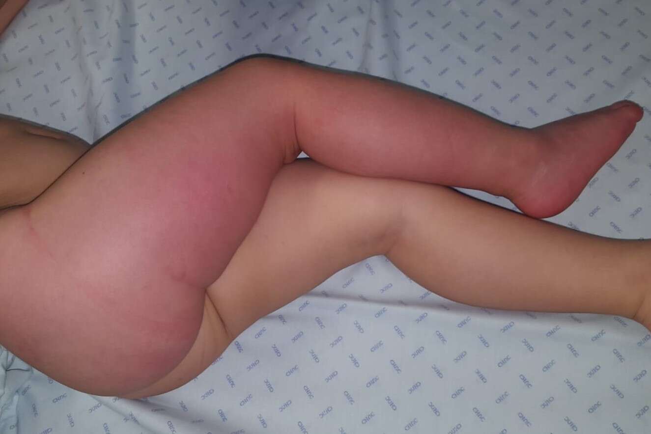
Case report: A 2-year old girl, without relevant personal or family medical history, presented with a lesion in the right lower limb with 2 months of evolution. No fever or other accompanying symptoms. Physical examination revealed a widespread pink patch with ill-defined borders, covering all the right lower limb with extension to the ipsilateral buttock and foot. Skin was lightly thickened, shiny and had loss of wrinkling, mainly on the foot area. Acute phase reagents were in the normal range and antinuclear antibodies and extractable nuclear antigens were negative. Serological evaluation was suggestive of recent infection by Borrelia burgdorferi with positive IgM antibodies (antigenic fractions of major proteins p41, p39, p18 and OspC) and negative IgG. Skin biopsy showed perivascular infiltrate with numerous eosinophils, mild sclerosis and hyalinization of collagen bundles at reticular dermis, concordant with localized scleroderma with deep involvement. Treatment was initiated with monthly intravenous methylprednisolone pulses (30 mg/kg), oral prednisolone (0,5 mg/kg/day) and subcutaneous methotrexate (18 mg/m2/week). It was also prescribed a 21-day cycle of amoxicillin. Two months after diagnosis, IgM and IgG antibodies for Borrelia burgdorferi were negative. Clinical evolution was favourable, with erythema resolution at 5 months and residual sclerosis in the foot at 15 months of treatment.
Conclusion: The role of borrelia infection as trigger of linear scleroderma currently has no scientific evidence, but cases describing the association continue to be reported. In this case, the occurrence of an extensive lesion in the inflammatory phase of scleroderma with simultaneous evidence of acute infection by Borrelia burgdorferi, lead to the hypothesis of a causal relationship between both entities.
Powered by Eventact EMS