
The Unyielding Dangers of Magnets: A Case Series
Background: Foreign body ingestion (FBI) is a common paediatric presentation, seen mostly in children between 6 months to 3 years. Multiple magnet ingestion is particularly dangerous as attraction of the magnets through bowel wall can lead to ischaemia, perforation, fistula, volvulus or necrosis.
Objective: We report two cases where older children presented to a district general hospital following the ingestion of multiple magnets.
Methods: Data was collected from electronic health record and patient interviews.
Results: Case 1: A 10-year-old girl presented with acute vomiting, fever, umbilical pain radiating to the right iliac fossa, a distended abdomen, and raised CRP (349). A CT scan (Figure 1) initially reported an acute perforated appendicitis with a calcified lobulated object in the caecum/appendix, likely an appendicolith.
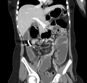
Paediatricians subsequently noticed her playing with magnetic balls that looked strikingly like the lobulated object found on imaging. She adamantly denied FBI. Following transfer to a tertiary centre, a laparotomy revealed loops of distal ileum stuck together by six magnets in the caecum, with bowel perforation. The magnets and 62 cm of ileum were excised, and an anastomosis formed.
Case 2: A 9-year-old boy accidentally ingested magnets while jumping on a trampoline with magnets in his mouth (as he enjoyed this sensation). Abdominal X-ray (Figure 2) revealed magnets clumped together.
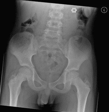
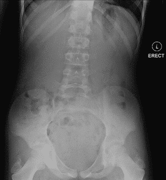
As this indicated low risk of perforation, he was monitored and repeat X-ray (Figure 3) confirmed safe passage of magnets.
Conclusion: FBI, though not uncommon, can be forgotten in older children. The first case demonstrates how unforthcoming even older children can be. Careful deduction allowed for the correct identification of a complex small bowel obstruction & perforation requiring specialist intervention. Case two highlights how the management of FBI can vary greatly depending on the history. Where this is uncertain, imaging can be essential for diagnosis and guiding treatment.
Powered by Eventact EMS