
Hepatic Inflammatory Pseudotumor Presenting in an 10-year-old Boy
2Department of Pediatric Surgery, Hospital de Braga, Portugal
3Great Osmond Street Institute of Child Health, University College London, Portugal
4The Life and Health Sciences Research Institute, Minho's University, Portugal
5Medical School, Minho's University, Portugal
Background: Inflammatory pseudotumors (IP) are a rare entity. Etiology is unknown and its course is also undefined ranging from spontaneous regression to metastatic dissemination. They mostly affect the lungs, but also occur in the liver and other organs. Most hepatic lesions present with abdominal pain and the treatment is still controversial.
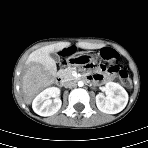
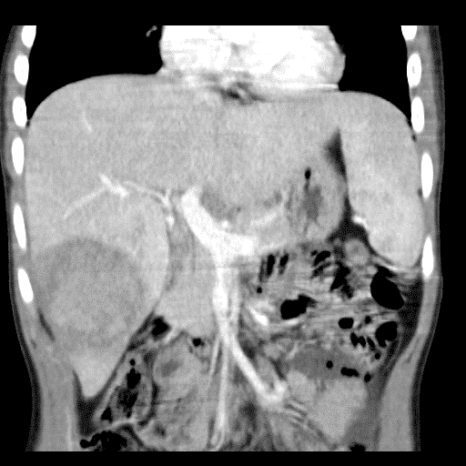
Description: A previously healthy 10-year-old boy presented with colicky abdominal pain in the right quadrants, intermittent fever of 1 month of evolution, sporadic vomiting and diarrhea. On physical examination he was ill-appearing; abdominal pain on palpation and a tender mass was found on right upper quadrant. Analytically, Hgb 9.7 mg/dl, leukocytes 23520/uL, sedimentation rate 102 mm/h, C-reactive protein 13.59 mg/dl, ferritin 444 ug/L, normal liver function tests except a slight increase in GGT. An infectious cause was excluded with extended blood work. Echographically: hepatomegaly with heterogeneous area on the right lobe. Abdominal CT was performed to clarify: heterogeneous 7.4x6cm nodular area in the VI/VII hepatic segments. He started Ceftriaxone, Clindamycin and Metronidazole IV improving his general condition after one week, but the mass was still palpable. A liver MRI was performed: persistence of 5.5x4.8cm nodule in the VI segment of the right lobe. A biopsy was performed to exclude malignancy and histology revealed an IP. He was discharged home after a 14-day course of IV antibiotics and started ibuprofen, 30mg/Kg/day. He was kept on conservative treatment and follow-up with periodic ultrasound scans: progressive reduction of the hepatic nodule until complete resolution shown in MRI, 11 months later.
Conclusion: IP are an extremely rare condition, presenting in most cases with abdominal pain. Having an unknown cause its best treatment, whether surgical or conservative, is a matter of disagreement. This case highlights a rare cause of abdominal pain, a common symptom in children, that was successfully managed by a conservative approach.
Powered by Eventact EMS