
Treatment of a Massive Venous Malformation of a Thigh with Localized Intravascular Coagulation
2Global Clinical Scholars Research Training, Department of Postgraduate Medical Education, Harvard Medical School, Boston, USA
Background: Patients with massive venous malformations (VMs) may be challenging to treat. The purpose of this presentation is to demonstrate the management of a difficult case of localized VM.
Methods: An egg-shaped subcutaneous tumor on the lateral side of the left thigh was noted in a term male newborn after an uneventful pregnancy. MRI at 2 months of age showed a tumor encompassing 2/3 of the left thigh. With age the size of the thigh was increasing, always following episodes of severe pain accompanied by fever. Angiography done at the age of 10 months showed no arteriovenous shunts. Two small arterial feeders were embolized without any clinical improvement. D-dimer was elevated to a max of 12250 ng/ml and fibrinogen decreased to 0.9 g/L at the age of 26 months when the biopsy was performed. VM was confirmed and low-molecular-weight heparin therapy initiated. This reduced patient’s pain and improved the coagulation status, but the boy was still unable to walk. At the age of 27 months he was referred to us. After a thorough analysis of accompanying MRIs a localized VM was assumed and surgical intervention suggested. After obtaining the written parental consent surgery was performed at the age of 28 months.
Results: Medial and lateral incisions along the complete thigh length were performed, neurovascular structures dissected, complete removal of the VM performed, and the thigh reconstructed. The patient recovered uneventfully. His coagulation parameters normalized and heparin therapy stopped at 3 months postoperatively. At 24 months follow-up he is disease free, without leg-length discrepancy and playing soccer with his friends.
Conclusion: Surgical approach may be curative and worthwhile trying in localized VMs even if they may appear gigantic or inoperable.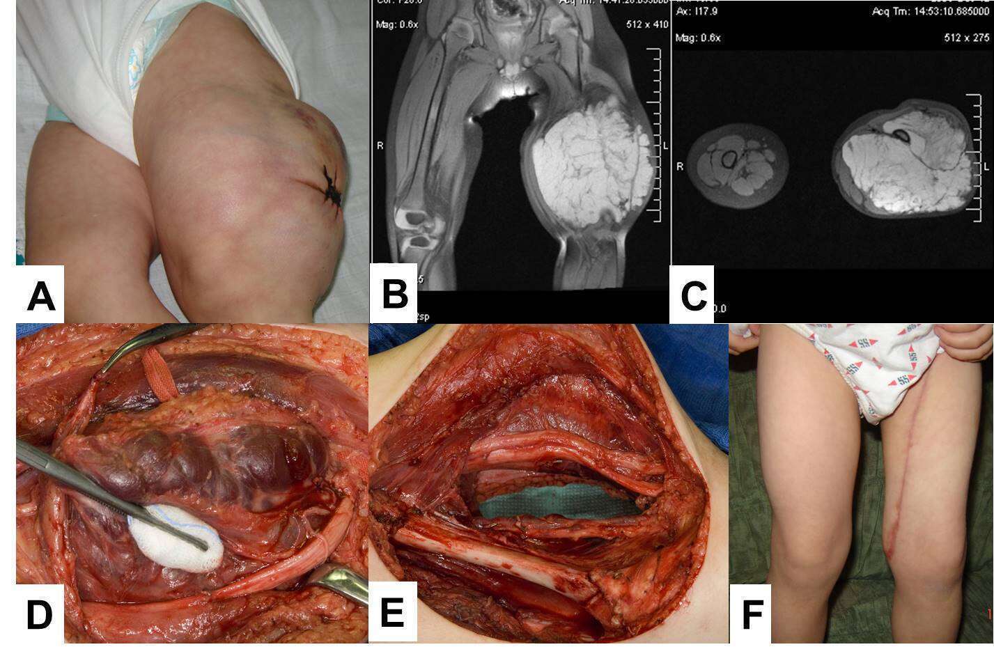
Powered by Eventact EMS