
Fetal Brain Biometry in Isolated Mega Cisterna Magna: MRI and US Study
2Department of Obstetrics and Gynecology, Sheba Medical Center, ישראל
3Sackler School of Medicine, Tel-Aviv University, ישראל
4Pediatric Neurology Unit, Sheba Medical Center, ישראל
Objective: To characterize the biometric parameters in ultrasound and brain MRI of fetuses with isolated mega cisterna magna (MCM).
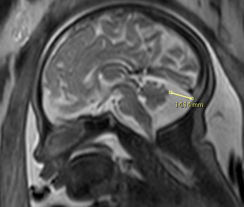
Methods: Cross-sectional historical cohort study conducted at a single tertiary medical center between 2011-2018. All fetuses underwent US and brain MRI scans. Matching analysis was performed according to gender and gestational age.
Results: The study included a total of 103 fetuses; 44 fetuses with isolated MCM in the study group, and a control group of 59 fetuses with normal CNS. The study group had larger biparietal diameter (BPD) (86 vs. 79.8 mm, p=0.001) and head circumference (HC) (318 vs. 292 mm, p<0.001) on ultrasound. On MRI, study group had larger occipitofrontal diameter (OFD) (99 vs. 92 mm, p<0.001) and BPD (77 vs. 72 mm, p<0.001). Male fetuses’ prevalence was higher in the study group (77.3 % vs. 47.5%). After matching 20 fetuses from each group, the study group had larger HC (310.1 versus 300.7 mm, p=0.029) and OFD (113.4 versus 108.3 mm, p=0.009) on ultrasound, and larger OFD (97.4 versus 94.6, p=0.013) on brain MRI.
Conclusions: Isolated MCM may be related to other large fetal CNS biometric measurements in both ultrasound and MRI and might be influenced by fetal gender.
.jpg)
.png)
.jpg)