
Anatomical Accuracy of the KODEX-EPD Novel 3D mapping system of the Left Atrium during Pulmonary Vein Isolation: a Correlation with Computer Tomography imaging.
2EPD, Philips, Israel
Background: A novel 3D mapping system (KODEX - EPD, EPD Solutions, Best, The Netherlands) enables catheter localization and real-time 3D cardiac mapping.
Objective. To evaluate left atrium (LA) anatomical mapping accuracy created by the EPD system during PVI compared with gold standard computed tomography (CT) images acquired from the same patients (pts) prior to the procedure.
Methods. In 15 consecutive pts who underwent PVI, 3D mapping of the LA was created on the EPD system using the Achieve catheter. PV, posterior wall, and appendage anatomy and diameters, were compared to the CT 3D reconstruction measured on the CARTO 3 system. Measurements were done independently by 2 physicians in each method. Linear correlation and agreement between CT and EPD measurements were assessed by Spearman correlation and Bland-Altman plot.
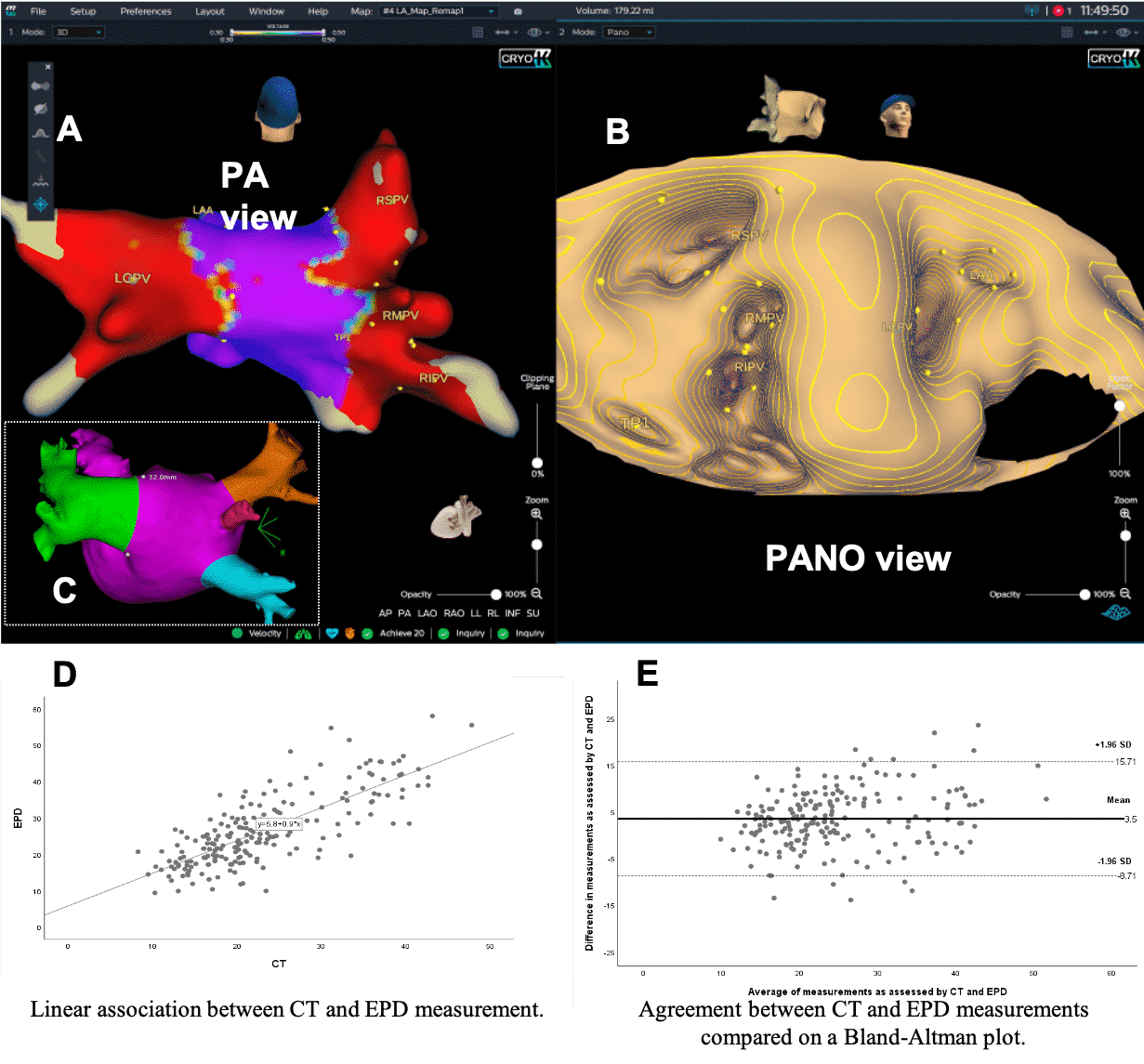
Results. Mean LA mapping time was 8+3.6 min. An EPD example map is shown in figure 1A, 1B, with corresponding CT map in 1C. In 4 (27%) and 3 (20%) pts disagreement regarding left common and right middle accessory vein anatomy was seen. Very high interobserver correlation was found for both EPD and CT measurements (Spearman-r 0.9). High correlation (r 0.75) was found between CT and EPD measurements. Bland-Altman plot method revealed that measurements assessed by EPD were slightly higher than those assessed by CT. Mean difference was 3.5mm, p<0.01(figure 1D, 1E).
Conclusion: The new EPD mapping system allows quick and accurate mapping of LA with high correlation to CT imaging. Some differences in left common and accessory right middle vein anatomy were seen.
Powered by Eventact EMS