
Automated Processing of Thermal Imaging to Detect
COVID-19 and Microvascular Dysfunction
2Sackler Faculty of Medicine, Tel Aviv University, Israel
3Faculty of Engineering, Tel Aviv University, Israel
4Internal Medicine B, Sheba Medical Center, Israel
5Medical Corps, Israel Defense Forces, Israel
6Department of Diagnostic Imaging, Sheba Medical Center, Israel
7Internal Medicine Wing and Hypertension Unit, Sheba Medical Center, Israel
8School of Medical Engineering, Afeka Tel Aviv Academic College of Engineering, Israel
9School of Electrical Engineering, Afeka Tel Aviv Academic College of Engineering, Israel
Background: Coronavirus disease 2019 (COVID-19) is associated with microvascular dysfunction. Non-invasive thermal imaging can hypothetically detect changes in perfusion, inflammation, and vascular injury. We sought to develop a new point-of-care, non-contact thermal imaging tool to detect COVID-19 by microvascular dysfunction, based on image processing algorithms and machine learning analysis.
Materials and Methods: We captured thermal images of the back of 101 individuals, with (n=62) and without (n=39) COVID-19, from two medical centers in Israel, using a portable thermal camera that connects directly to smartphones. We developed new image processing algorithms that automatically extract multiple texture and shape features of the thermal images (Figure 1A). We then evaluated the ability of our thermal features to detect COVID-19 and systemic changes of heat distribution associated with microvascular disease. We also assessed correlations between thermal imaging to conventional biomarkers and chest X-ray (CXR).
Results: Our novel image processing algorithms achieved up to 92% sensitivity in detecting COVID-19 with an area under the curve of 0.85 (95% CI: 0.78, 0.93; p < 0.01). Systemic alterations in blood flow associated with vascular disease were observed across the entire back. Thermal imaging scores were inversely correlated with clinical variables associated with COVID-19 disease progression, including blood oxygen saturation, C- reactive protein, and D-dimer. The thermal imaging findings were not correlated with the results of CXR.
Conclusions: We show, for the first time, that a hand-held thermal imaging device can be used to detect COVID-19. Non-invasive thermal imaging could be used to screen for COVID-19 in out-of-hospital settings, especially in low-income regions with limited imaging resources. Moreover, thermal imaging might detect micro-angiopathies and endothelial dysfunction in patients with COVID-19 and could possibly improve risk stratification of infected individuals (Figure 1B).
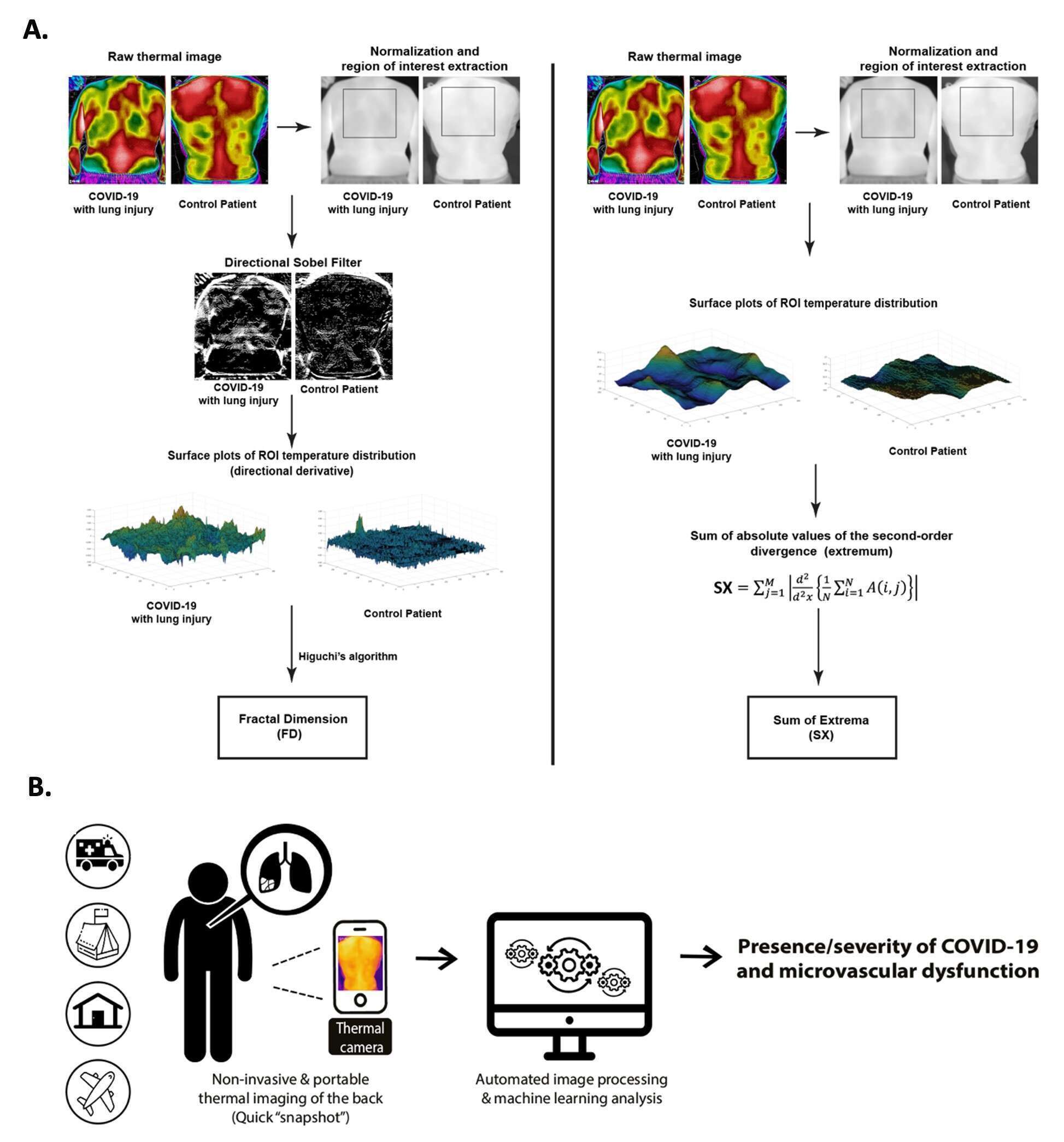
Figure 1: A. Representative steps of our thermal image processing algorithms. B. A schematic illustration of the research design and potential impact.
Powered by Eventact EMS