HIPAK Annual Meeting 2022

Tissue Damage Markers as Predictors of non-response to Anti-Tumor Necrosis Factor-α Agents in Children with Crohn’s Disease

טל קוזקוב 1 Alex Krauthammer 1,2 Sophia Fried 1 Adi Har-Zahav 1 Raanan Shamir 1,2 Amit Asa 3 Orith Waisbourd-Zinman 1,2
1Pediatric Gastroenterology, Nutrition and Liver Diseases, Schneider Children’s Medical Center, ישראל
2Sackler Faculty of Medicine, Tel-Aviv University, ישראל
3the Juliet Keidan Institute of Paediatric Gastroenterology and Nutrition, Shaare Zedek Medical Center, ישראל

Objectives and Aims: Current therapeutic strategies in patients with severe Crohn’s disease (CD) is anti-tumor necrosis factor-α (anti TNF-α) agents. While some patients show excellent response to treatment, others, show no or only partial response. Little is known about immunohistochemical tissue markers that can assist predicting patient’s response to those agents. The aim of our study was to identify immunohistochemical predictors of response to anti TNF-α agents via biopsies from the terminal ileum in pediatric CD patients.

Methods: We retrospectively reviewed medical records of pediatric patients with CD, who were treated with anti TNF-α treatment induction. Patients were divided to three equal groups (full, partial, and non-responders) according to their clinical, mucosal and laboratory response to treatment. Nine tissue inflammatory markers were evaluated, and immunofluorescence (IF) analyses was preformed to identify, quantify, and compare the differences between the groups, using biopsies from the terminal ileum.

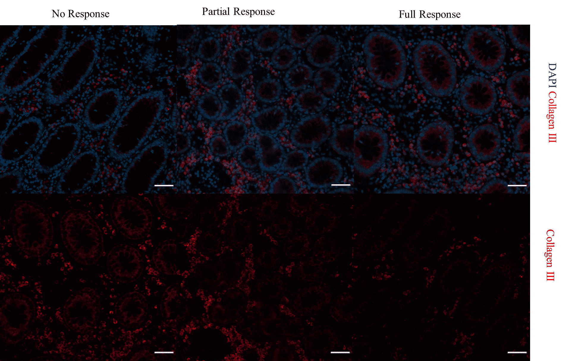
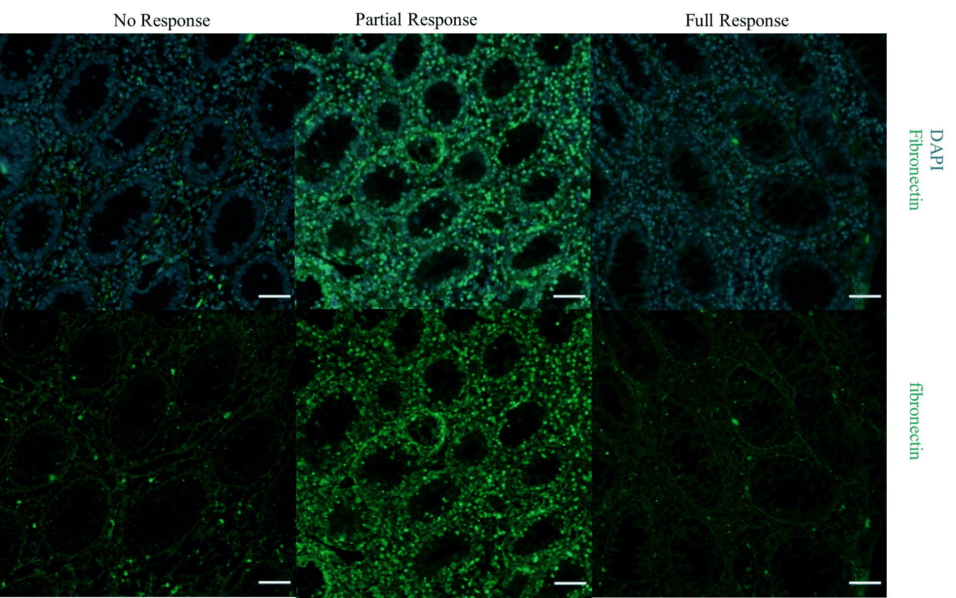
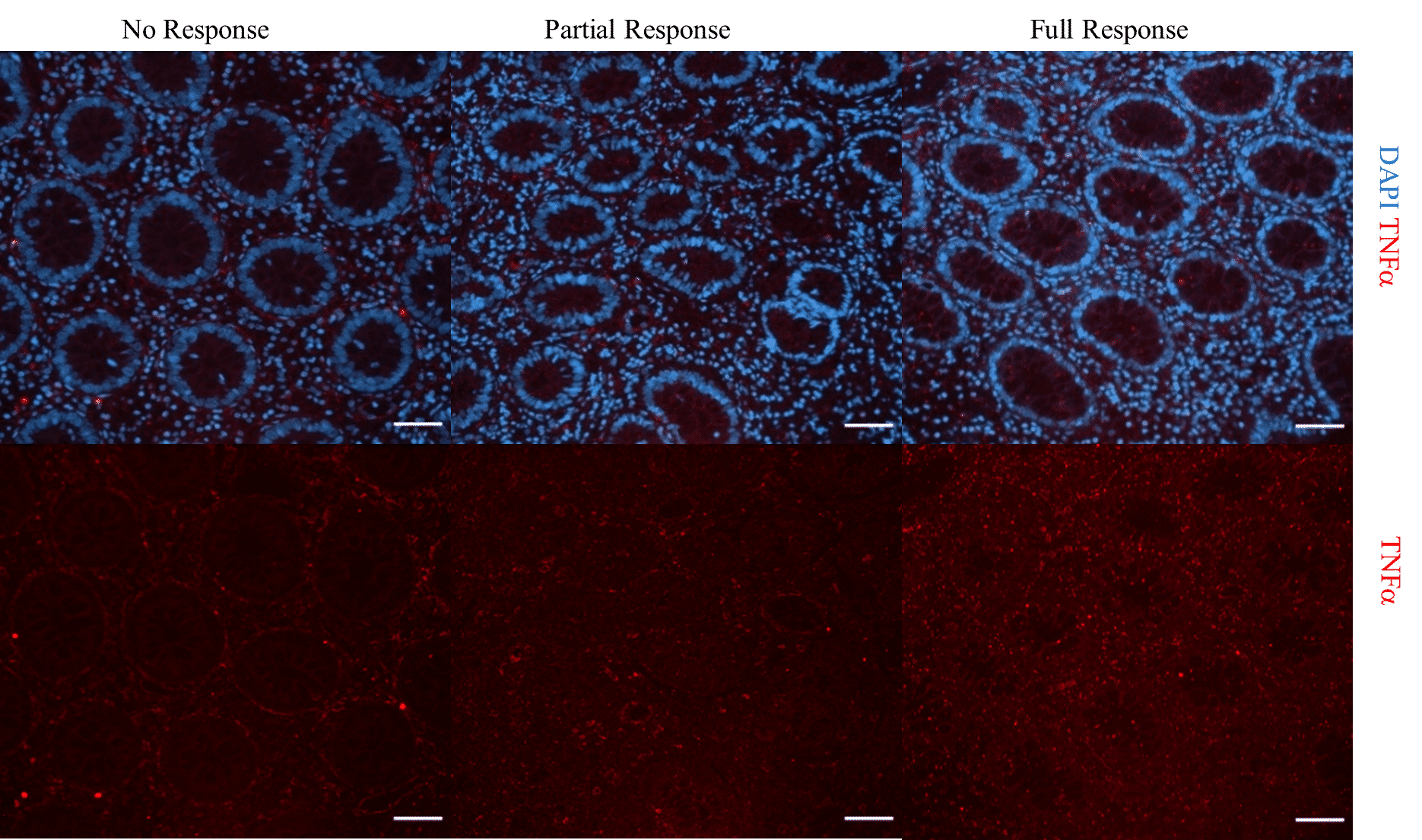
Results: Twenty-seven patients were included in this study (n=9, each group). Three out of nine evaluated markers showed difference in IF intensity (Fig.1). TNF-α stain was significantly more pronounced in the responder`s group and predicted response with a significant statistical correlation (TNF-α, p<0.05) while collagenIII and fibronectin were more pronounced in the non-responder`s group (p<0.05, each). These markers could not differentiate between FR and PR.

Conclusion: In pediatric CD patients, increased IF stain of fibronectin and collagenIII on biopsies prior to initiation of anti TNF-α may predict unresponsiveness to anti TNF-α treatment, while increased stain of TNF-α, may predict response.Figure 1- Terminal ileum biopsies of CD patients stained for TNF-, collagen III and fibronectin in the three groups of patients - NR (no response) PR (partial response) and FR (full response) may represent predictors of the treatment response. A – TNF- in red, there was markedly increased expression in the FR group. B- collagen III in red and C- fibronectin in green were expressed more prominent in the NR group in a diffuse manner.  Scale bar 50 µmFigure 1- Terminal ileum biopsies of CD patients stained for TNF-, collagen III and fibronectin in the three groups of patients - NR (no response) PR (partial response) and FR (full response) may represent predictors of the treatment response. A – TNF- in red, there was markedly increased expression in the FR group. B- collagen III in red and C- fibronectin in green were expressed more prominent in the NR group in a diffuse manner.  Scale bar 50 µmFigure 1- Terminal ileum biopsies of CD patients stained for TNF-, collagen III and fibronectin in the three groups of patients - NR (no response) PR (partial response) and FR (full response) may represent predictors of the treatment response. A – TNF- in red, there was markedly increased expression in the FR group. B- collagen III in red and C- fibronectin in green were expressed more prominent in the NR group in a diffuse manner.  Scale bar 50 µm

Figure 1- Terminal ileum biopsies of CD patients stained for TNF-, collagen III and fibronectin in the three groups of patients - NR (no response) PR (partial response) and FR (full response) may represent predictors of the treatment response. A – TNF- in red, there was markedly increased expression in the FR group. B- collagen III in red and C- fibronectin in green were expressed more prominent in the NR group in a diffuse manner.  Scale bar 50 µm