")
GOLDEN VATERITE AS A MESOSCOPIC METAMATERIAL FOR BIOPHOTONIC APPLICATIONS
Mesoscopic photonic systems with tailored optical responses have great potential to open new frontiers in implantable biomedical devices. However, biocompatibility is typically a problem, as engineering of optical properties often calls for using toxic compounds and chemicals, unsuitable for in vivo applications. Here, a unique approach to biofriendly delivery of optical resonances is demonstrated. It is shown that the controllable infusion of gold nano seeds into polycrystalline sub-micrometer vaterite spherulites gives rise to a variety of electric and magnetic Mie resonances, producing a tuneable mesoscopic optical metamaterial. The 3D reconstruction of the spherulites demonstrates the capability of controllable gold loading with volumetric filling factors exceeding 28%. Owing to the biocompatibility of the constitutive elements, “golden vaterite” paves the way to introduce designer-made Mie resonances to cutting-edge biophotonic applications. This concept is exemplified by showing efficient laser heating of gold-filled vaterite spherulites at red and near-infrared wavelengths, highly desirable in photothermal therapy, and photoacoustic tomography.
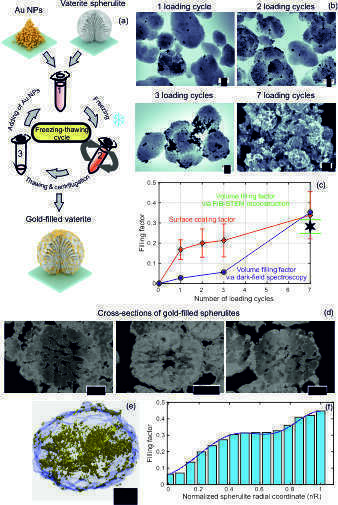
Schematics of infusing gold nanoparticles (Au NPs) into vaterite spherulites via freezing-induced loading and b–f) characterization of the resulting gold-filled spherulites. The variations in the number of loading cycles allows control of the filling factor. SEM images for one, two, and three loading cycles in (b) are presented in the inverse color scheme to highlight the sparse distributions of gold nano inclusions (black color) whereas the case of seven loading cycles is shown in the normal color scheme. The surface coating and volume filling factors as functions of the number of loading cycles are shown in (c). The mean and the standard deviation are obtained by the statistical analysis of a large number of spherulites in the high resolution SEM and STEM images. Also, the volume filling factor is evaluated by numerical fitting to the dark-field scattering spectra, shown in Figure 2. The cross-sections of vaterite spherulites loaded with 7 cycles in (d) are obtained by etching with the focused ion beam and then imaged via scanning transmission electron microscopy. SEM and STEM images were taken before annealing. The 3D reconstruction in (e) is performed by processing a z-stack of STEM images in MATLAB. The spatial distribution for Au NPs from the reconstruction in (e) is shown in (f). The distribution is fitted by a sum of two Gaussian functions