")
NOVEL FERROMAGNETIC EuInAs/ InAsSb NANOWIRES BY MBE
2Department of Chemical Research Support, Weizmann Institute of Science, Rehovot, Israel
3Department of Physics and Institute of Nanotechnology and Advanced Materials, Bar-Ilan University, Ramat-Gan, Israel
EuIn2As2 is predicted to be a magnetic topological-crystalline insulator. It consists of alternately stacked divalent Eu (Eu2+) and In2As2 layers along the c-axis [1,2]. Bulk single crystals of EuIn2As2 have been synthesized via a flux method with a by-product which includes trivalent Eu (Eu3+). Here, we report novel ferromagnetic EuInAs on InAsSb nanowires by molecular beam epitaxy (MBE). MBE growth can provide various binary and ternary composition nanowires that can support hybrid nanoscale devices such as superconductor-semiconductor [3] or ferromagnetic insulator-semiconductor [4] materials with a geometrical variety.
Wurtzite (WZ) structure InAs nanowires were first grown by the gold-assisted vapor–liquid–solid (VLS) mechanism on epi-ready (001) InAs substrates serving as stems followed by axial growth of Zinc blende (ZB) structure InAs0.9Sb0.1 (Sb 5%)[5]. Incorporation of Eu turns out to result in an EuInAs shell (or partial shell) covering the nanowires, in which a unique ZB like crystal structure was observed. Interestingly, Eu ions induce the inversion domain boundary (IDB) between As atoms. Separated Eu ion layers are distributed with rhombic patterns throughout the shell ZB InAs structure, which is compatible with the InAsSb ZB structure on the one hand and expected to have ferromagnetic properties on the other hand. EuInAs shell coating was initially performed on reclined InAs nanowires. The WZ InAs has a rough EuInAs surface, whereas the so-called stalactite (vertical) InAs nanowire which is formed at the intersection of two WZ InAs nanowires [6] has a smooth and uniform surface Scanning electron microscopy (SEM) and high-resolution transmission electron microscopy (HR-TEM) were performed to study the morphology and structure of EuInAs nanowires in detail, respectively. High-angle annular dark-field scanning TEM (HAADF-STEM) and energy-dispersive X-ray spectroscopy (EDS) mapping provided extensive information regarding the atomic coordination and composition of the distinctive crystalline EuInAs structure. Scanning superconducting quantum interference device (SQUID) is used to study the magnetic properties of EuInAs structure.
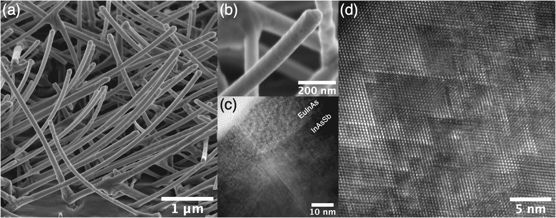
Figure 1 (a) and (b) Bird’s-eye view SEM images of InAsSb–EuInAs core–partial shell nanowires. (c) an HR-TEM image of the interface between core and shell. (d) Rhombic pattern signifies the inversion domain boundaries induced by Eu ions.
[1] A. M. Goforth, P. Klavins, J. C. Fettinger, and S. M. Kauzlarich, Inorg. Chem. 47, 11048 (2008).
[2] S. X. M. Riverolles, T. V. Trevisan, B. Kuthanazhi, T. W. Heitmann, F. Ye, D. C. Johnston, S. L. Bud’ko, D. H. Ryan, P. C. Canifield, A. Kreyssig, A. Vishwanath, R. J. McQueeney, L. -L. Wang, P. P. Orth, and B. G. Ueland, Nat. Commun. 12, 999 (2021).
[3] J. -H. Kang, A. Grivnin, E. Bor, J. Reiner, N. Avraham, Y. Ronen, Y. Cohen, P. Kacman, H. Shtrikman, and H. Beidenkopf, Nano Lett. 17, 7520 (2017).
[4] Y. Liu, S. Vaitiekėnas, S. Martí-Sáchez, C. Koch, S. Hart, Z. Cui, T. Kanne, S. A. Khan, R. Tanta, S. Upadhyay, and M. E. Cachaza, C. M. Marcus, J. Arbiol, K. A. Moler, and P. Krogstrup, Nano Lett. 20, 456 (2020).
[5] T. Xu, K. A. Dick, S. Plissard, T. H. Nguyen, Y. Makoudi, M. Berthe, J. -P. Nys, X. Wallart, B. Grandidier and P. Caroff, Nanotechnology 23, 095702 (2012).
[6] J. -H. Kang, M. Galicka, P. Kacman, and H. Shtrikman, Nano Lett. 17, 531 (2016).