")
HYBRID-PIXEL DETECTORS FOR TEM BY DECTRIS
25 years of advances in aberration correction have clearly shifted the TEM characterization limits from the electron optics to other factors, among them the electron detectors. Recent improvements on electron detection technology clearly impact the TEM characterization on both Materials Sciences and Life Sciences, particularly when beam-sensitive samples are involved [1]. Hybrid-pixel detector (HPD) [2] is one of the approaches to directly detect and count electrons, with the distinctive advantage of flexible design with respect to the sensor material and electronics for optimization to different applications.
Building from its successful HPD technology for X-rays detectors, recently DECTRIS optimized its design to enable the precise detection of electrons. Among the required adaptations were the determination of optimal threshold values for counting electrons with zero read-out noise within a wide energy range and the retrigger technology fine-tuning to allow counting from 1 to 107 counts/pixel/second [3].
These characteristics indicate that HPDs can contribute to a variety of TEM applications, including low-dose imaging with single-electron sensitivity, 4D-STEM acquisition with simultaneous high dynamic range and frame rate (2,250 fps with full read-out and 18,000 fps with a reduced area), and flexible EELS collection – from zero-loss peak (ZLP) to zero-noise core-loss (CL) region included in the same acquisition range (Figures 1 and 2 – adapted from reference [3]).
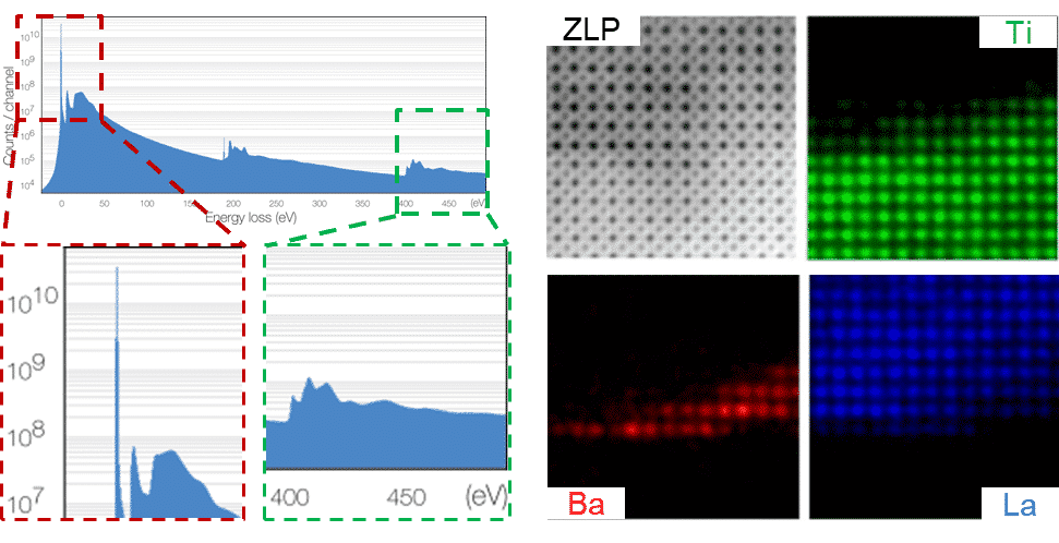
Fig. 1. (left) EELS spectrum of h-BN at 60kV. Simultaneous ZLP and CL collection without saturation across 6 orders of magnitude. (right) Spectrum imaging of STO/BTO/LSMO. Flexible elemental mapping with multipass EELS allowed by zero-readout noise.
References:
[1] A. R. Faruqi et al., Nucl. Inst. Methods Phys. Res., A 878, 180-190 (2018).
[2] N. Bochenek et al., IEEE Trans Nucl Sci 65 (6), 1285-1291 (2018).
[3] B. Plotkin-Swing et al., Ultramicroscopy 217, 23-30 (2020).