Imaging the electric field in bulk ferroelectrics
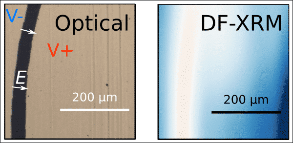
Emergent properties can be measured extrinsically using electrodes, imaged at surfaces using scanning probe microscopy (SPM) or in lamella using transmission electron microscopy (TEM). Despite tremendous advances in SPM and TEM over the past decades, these techniques cannot penetrate the bulk. X-ray-based techniques can penetrate through the material and are well suited for investigation of the bulk, but investigation of polarization with X-rays remains in its infancy. Multiple X-ray techniques are now able to image strain and orientation deep in the bulk, and dark-field X-ray microscopy (DF-XRM) has previously been used to image domains deep in the bulk of ferroelectrics [1]. Here, we present our efforts to image electric-field induced polarization using DF-XRM, in addition to domains and strain. This work will enable deeper analysis of interplay between strain, stress and polarization in the 3D bulk of the material, as well as imaging of exotic topological structures deep inside the sample.
In this talk, we will provide proof-of-concept images of the electric field acquired with DF-XRM. Our results are backed by a theoretical exploration of the contrast mechanism used to image polarization, and simulations of realistic experimental conditions. Experimental results from DF-XRM convolute information on strain, orientation and polarization, which makes analysis non-trivial. The theoretical work we present here will show how contrast from an applied electric field, strain and orientation can be separated, and is therefore relevant in any work where electric field is applied to a sample in DF-XRM.
[1] Simons, Hugh, et al. "Long-range symmetry breaking in embedded ferroelectrics." Nature materials 17.9 (2018): 814-819.
Powered by Eventact EMS
