The 72nd Annual Conference of the Israel Heart Society in association with the Israel Society of Cardiothoracic Surgery

Cardiac Tamponade Complicating Transcatheter Aortic Valve Replacement: Insights from a Single-Center Registry

Ibrahim Naoum Amnon Eitan Hussein Sliman Avinoam Shiran Salim Adawi Ihab Asmer Keren Zissman Ronen Jaffe
Department of Cardiology, Lady Davis Carmel Medical Center, Technion-Israel Institute of Technology, Israel


Background: Cardiac tamponade complicating transcatheter aortic valve replacement (TAVR) typically results from right ventricular (RV) injury induced by a pacemaker electrode, left ventricular (LV) injury induced by guidewires and catheters used during the procedure and rupture of the aortic annulus during valve implantation.
Methods: We retrospectively analyzed our institutional TAVR database to gain mechanistic insights relating to this complication.
Results: A total of 1,247 TAVR procedures were performed between 2010-2024. Cardiac tamponade complicated 21 (1.7%) of these procedures. There was a non-significant reduction in occurrence of tamponade (1.9% among the first 623 cases vs. 1.4% among the subsequent 624 cases, p=0.44). Tamponade was caused by LV perforation in 10 cases (48%), pacemaker-induced RV perforation in 8 cases (38%) and annular rupture in 3 cases (14%). We identified 2 mechanisms causing LV perforation: the stiff guidewire used for valve delivery caused myocardial injury in 7 cases. In the 3 remaining cases, LV perforation occurred prior to insertion of the stiff guidewire and was attributed to insertion of soft guidewires. No additional such cases occurred following implementation of a protocol for meticulous guidewire insertion into the LV. Pericardiocentesis was performed in 20 patients with tamponade and cardiac surgery in 13. Nine patients (43%) died during the index hospitalization. Mortality did not differ between cases with RV perforation and left sided perforation.
Conclusions: Peri-procedural cardiac tamponade during TAVR may be caused by various mechanisms. Careful guidewire manipulation may decrease occurrence of LV perforation.
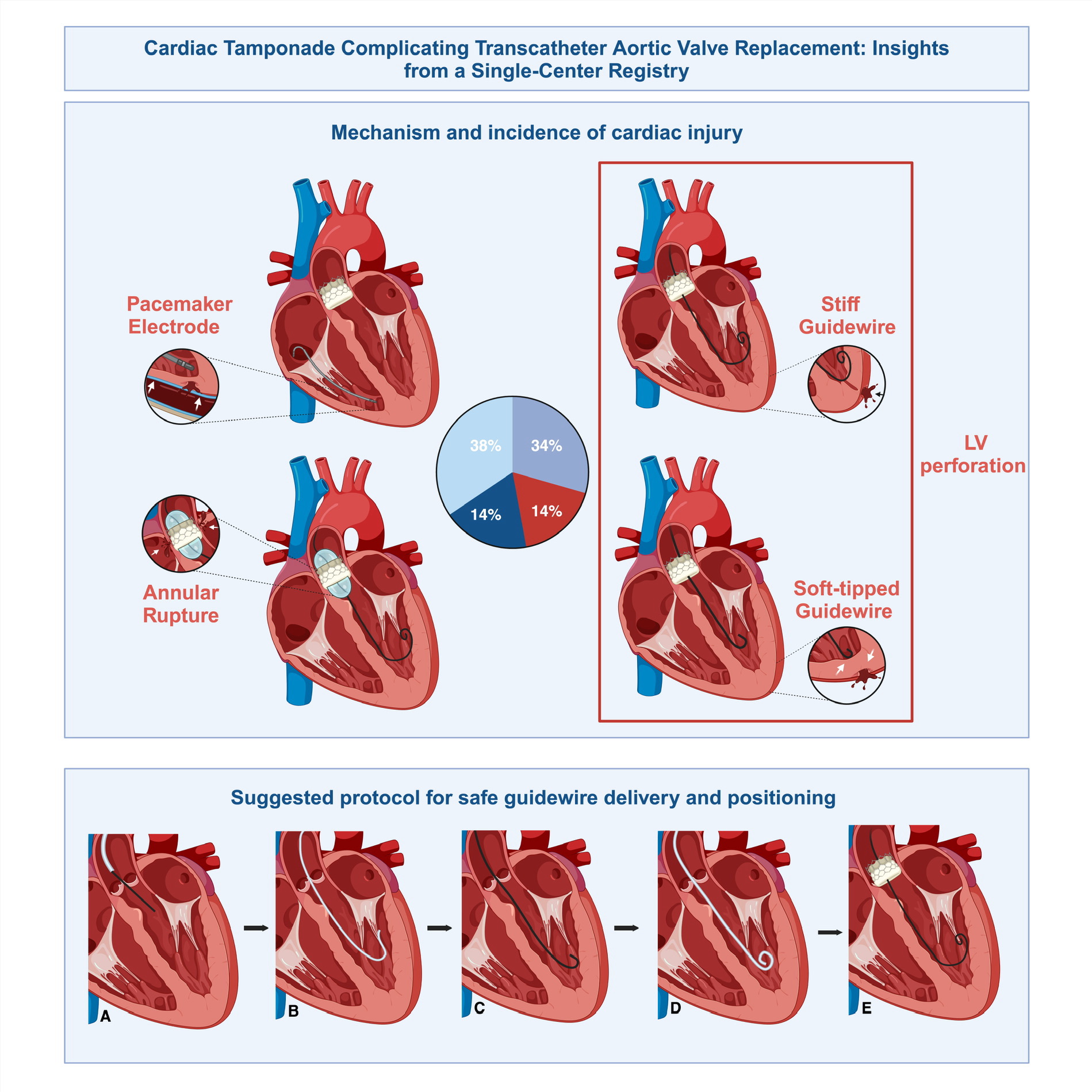
Central Illustration: Cardiac tamponade during transcatheter aortic valve replacement (TAVR): Upper panel: Mechanism and incidence of cardiac injury among patients with tamponade. Lower panel: Suggested protocol for safe guidewire positioning within the left ventricle: A) Straight-tipped 0.035” soft guidewire inserted via an Amplatz-Left (AL) catheter across the stenosed aortic valve, B) AL catheter advanced over the straight-tipped guidewire, positioned in left ventricle (LV). Blood backflow from its rear end is verified, C) J-tipped soft guidewire advanced via the AL catheter and positioned within the LV, D) Pigtail catheter carefully advanced over the J-tipped guidewire and positioned within the LV. Gentle manual LV angiogram confirms pigtail catheter optimal location, E) Stiff guidewire advanced via pigtail catheter, positioned within the LV cavity and used for delivering the transcatheter heart valve to the implantation site.

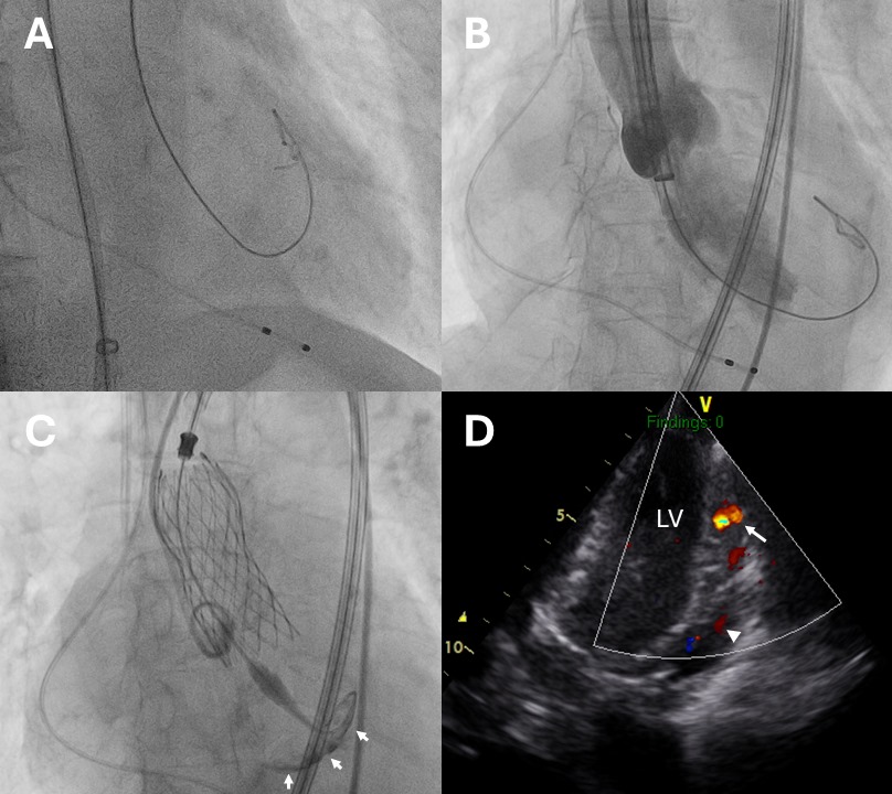
Left ventricle (LV) wire perforation: A) Fluoroscopic image of the stiff guidewire, presumably located within the LV cavity (right anterior oblique view), B) Aortogram during valve positioning: regurgitated contrast media filling the LV cavity, tip of guidewire located outside of  LV (left anterior oblique view), C) Fluoroscopic image following valve deployment-contrast media staining at LV apex (arrows), D) Color Doppler echocardiography (apical view) showing blood flow (arrow) from the LV to the pericardial space (arrowhead).

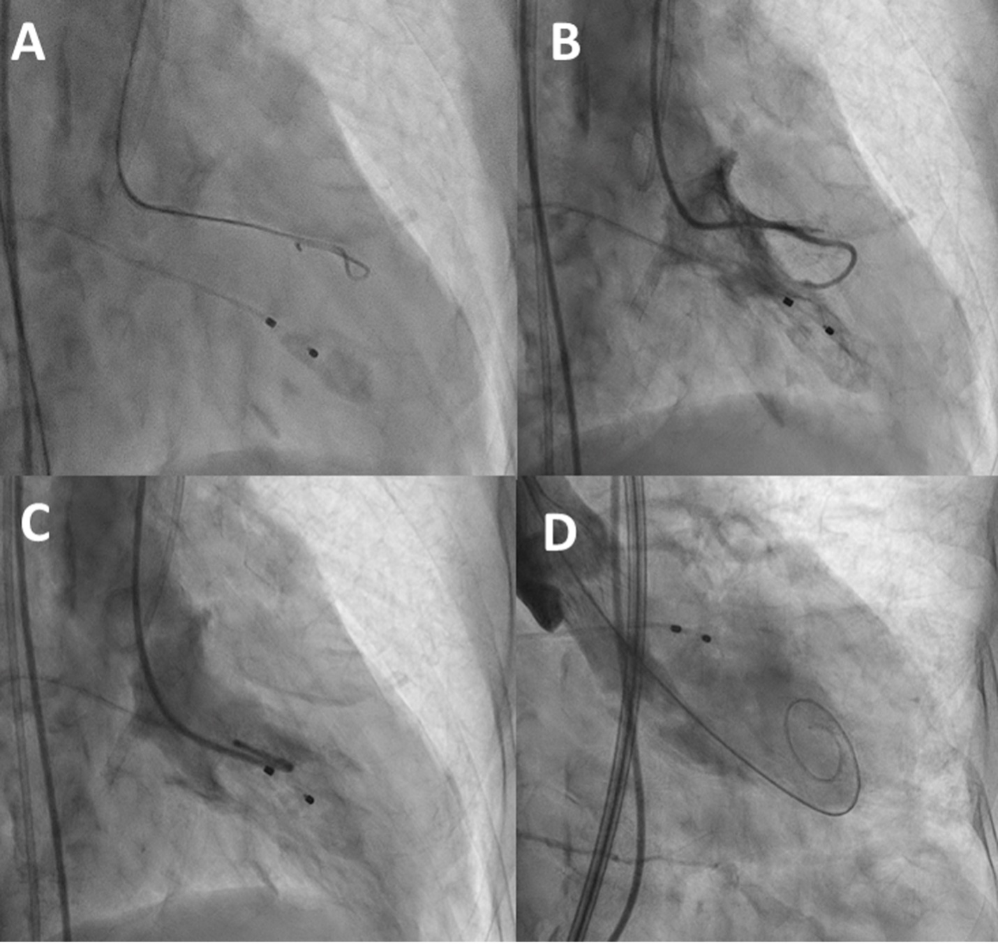
Left ventriculography for guiding guidewire position: A) Fluoroscopic image of initial guidewire position following insertion across the aortic valve, B) Ventriculography using gentle manual injection, showing tip of catheter behind a muscular trabeculae C) Repeat ventriculography following catheter repositioning within the LV cavity, D) Guidewire located within LV apex during valve deployment.