Assessment of Aortic Insufficiency Severity Using Color-Doppler in Mitral Short-Axis View by Measuring the Perimeter and Area of Diastolic Blood Flow Anterior to Mitral Leaflet
2Heart Center, Sheba Medical Center, Israel
Introduction Assessing the severity of aortic valve insufficiency (AI) through echocardiography can be challenging. While guidelines suggest various evaluation methods, they all require optimal imaging of the valve, the left ventricular outflow tract, and the characterization of the regurgitant jet. Quantitative Doppler measurements can sometimes lead to inaccurate estimations of severity based on the acuity of AI development. This issue is particularly pronounced when assessing artificial valves or paravalvular leaks. We aimed to correlate the size of the AI regurgitant jet, represented in Color Doppler Mitral short axis (MSAX) views, with the severity of AI.
Methods We analyzed our Echocardiography lab registry for patients with >moderate aortic regurgitation compared to those with moderate or lesser degrees. AI severity was evaluated by calculating the vena contracta diameter (AI_VC) and identifying the presence of holodiastolic flow reversal in the descending aorta. A patient was classified as having >moderate AI if either AI_VC was >5mm or holodiastolic flow reversal was observed. Left ventricular outflow dimension (LVOTd) was measured, and the AI_VC/LVOTd ratio was calculated. Additionally, the MSAX AI area and perimeter were measured and correlated with guideline-directed AI severity. Test performance was assessed using ROC testing.
Results 23 patients were classified as having AI > moderate, while 55 had AI ≤ moderate. The average age of patients with AI ≤ moderate was 73±15 years, compared to 65±24 years for those with >moderate AI (p=0.022). Table show echo findings. MSAX AI perimeter >3.14cm demonstrated 100% sensitivity and 73% specificity for >moderate AI, with an area under the curve of 91% (p<0.001).
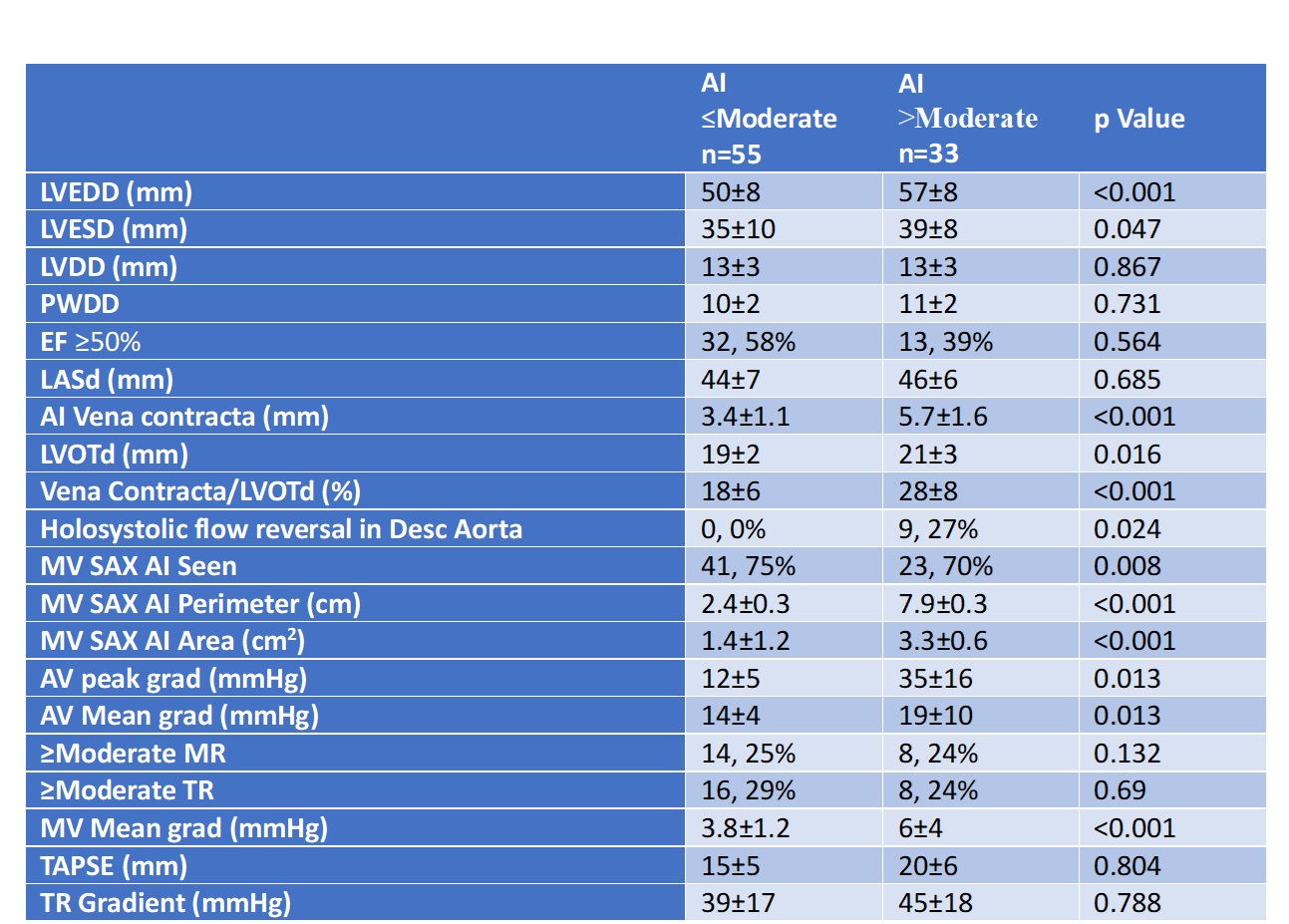
Conclusion The MSAX AI perimeter shows promise as a diagnostic tool for significant AI. Further research is needed to evaluate its effectiveness in complex clinical scenarios, such as paravalvular leaks after aortic valve implantation, where traditional metrics often fail due to imaging limitations.
israelheart@ortra.com מייל לשאלות