Inverse Correlation Between Coronary Lumen Volume to Left Ventricular Mass Ratio and Myocardial Fibrosis in Patients with Hypertrophic Cardiomyopathy
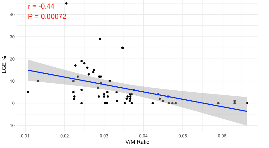
Introduction: In hypertrophic cardiomyopathy (HCM)y, inadequate myocardial perfusion can contribute to myocardial fibrosis. The coronary lumen volume (V) to left ventricular mass (M) ratio (V/M ratio) may serve as a surrogate marker for myocardial perfusion relative to myocardial mass. This study aims to evaluate the relationship between the V/M ratio determined by computed tomography (CT) and late gadolinium enhancement (LGE) determined by cardiac magnetic resonance imaging (MRI), a marker of myocardial fibrosis, in patients with HCM.
Methods: This retrospective cohort study included consecutive patients with HCM who underwent CT and MRI scans in the Rabin Medical Center. Patients with obstructive coronary artery disease were excluded. The entire epicardial coronary tree was automatically detected to calculate CBV, left ventricle was segmented to calculate LVM, and the V/M ratio was calculated and correlated with the extent of LGE on MRI. Pearson’s correlation analysis was used to assess this relationship.
Results: Our study included 57 patients with HCM. The median age was 59.6 years, and 71% were males. Cardiac MRI was used to assess several parameters, including LV mass (median 17.5 mm, IQR 11.75–24.25 mm), septal thickness (median 16 mm, IQR 11.5–20 mm), left ventricular ejection fraction (LVEF) (median 68.5%, IQR 63.25–73%), and LGE (median 4%, IQR 1-12%).
Conclusions: In patients with hypertrophic CMP a mismatch between coronary lumen volume and left ventricular mass is associated with increased myocardial fibrosis. This relationship emphasizes the V/M ratio’s potential as a non-invasive biomarker for identifying myocardial fibrosis and stratifying risk in this patient population.
israelheart@ortra.com מייל לשאלות