Atrial fibrosis in left atrial arrhythmias: results of comparative analysis by low voltage area mapping
2Biosense-Webster, Biosense-Webster, Israel
Background
Atrial fibrosis plays a significant role in both atrial fibrillation (AF) and atypical (left) atrial flutter (AAFL), but the differences in the substrate of these arrhythmias are not established. Low voltage areas (LVA) measurements on 3D mapping are a potential surrogate for local fibrosis.
Objective
To examine whether AAFL patients would have a higher percentage of atrial fibrosis than Persistent AF patients, using LVA quantification.
Methods
This retrospective single-center study included consecutive patients who underwent ablation for persistent AF and AAFL using the Biosense Webster CARTO mapping system from 2023 to 2024 and had bipolar voltage maps amenable to analysis. Measurements of very low voltage (0-0.2mv), low voltage (0-0.5mv), and normal (>0.5mv) areas were made using the CARTONET`s (Biosense Webster) voltage histogram. Detailed voltage analysis was performed for the entire left atrium and specifically segmented for anterior and posterior walls. Statistical comparisons were conducted using the Mann-Whitney U test.
Results
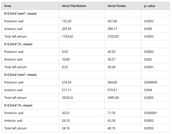
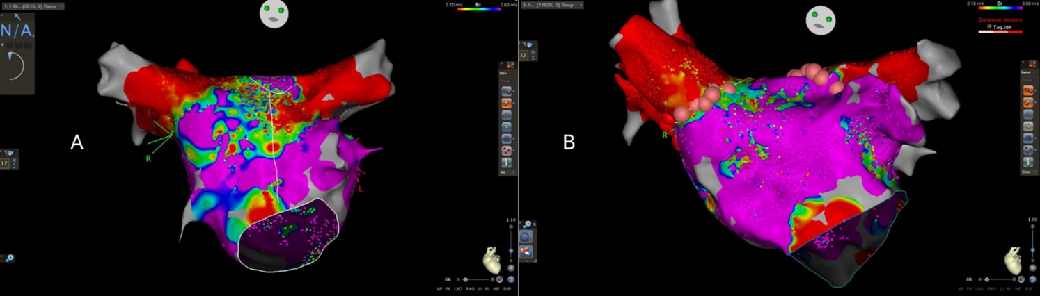
Eighteen AAFL patients and twenty AF patients were included in the study. The mean age was 66 and 70 (p=0.32)in the AAFL and AF groups. The rate of left atrial enlargement (moderate or higher) was 47.0% and 61.1% (p=0.40), respectively. Preserved left ventricular systolic function rate was 83.3% and 62.5% (p=0.17), respectively. Mean left atrial low voltage and mean very-low voltage areas, were significantly higher in AAFL, comparing to AF with 5685.86mm2 vs. 2928.25mm2 (p=0.0005) and 3102.82mm2 vs. 1163.42mm2 (p=0.0002) respectively. Significantly larger low and very low-voltage areas were also measured in a separate analysis of the anterior and posterior walls.
Conclusion
AAFL is associated with significantly more extensive areas of left atrial fibrosis than persistent AF. This finding may represent a more progressive atrial disease in AAFL that gives rise to reentry circuits.
israelheart@ortra.com מייל לשאלות