The relationship between left atrial electroanatomical voltage mapping and CMR-based fibrosis for enhanced preplanning in atrial fibrillation ablation
2Faculty of Medicine, Hebrew University of Jerusalem, Israel
Introduction: Cardiac magnetic resonance imaging-based late gadolinium enhancement (LGE-CMR) of left atrial (LA) fibrosis distribution can be valuable for preprocedural planning, patient selection and real-time guidance during atrial fibrillation (AF) ablation.
Aim: To evaluate the accuracy of LGE-CMR-based LA fibrosis (CMR-fib) among ablation-naive patients compared to electroanatomical-mapping (EAM-fib).
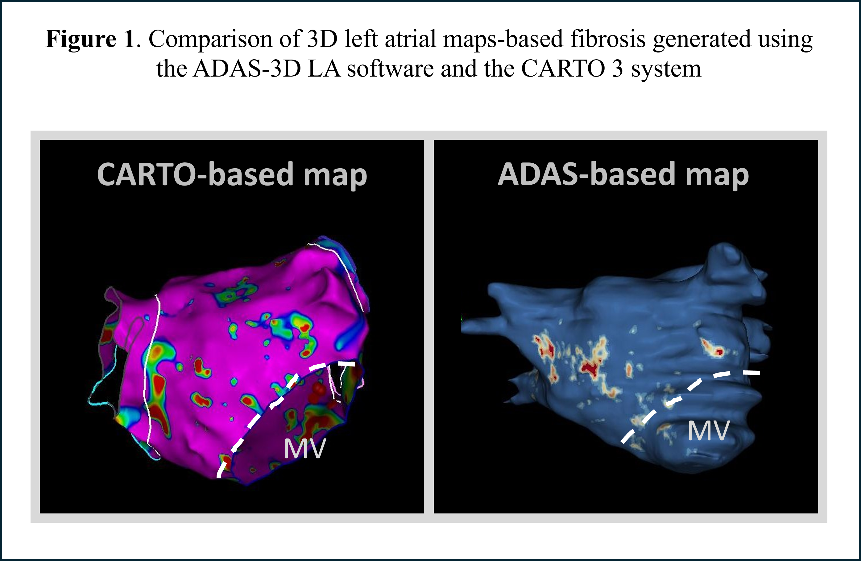
Methods: Patients who underwent CARTO-based mapping with preprocedural LGE-CMR were included. 3D substrate atrial maps were generated using the ADAS-3D-LA software and the CARTO 3 system with a fibrosis threshold of 1.32 and 0.1mV, respectively (Figure 1). The two maps were evaluated for fibrosis through anatomically synchronized, quantitative point-by-point comparison, with EAM used as the gold standard.
Results: The study included 41,543 points acquired from 24 patients (aged 64±11 yrs; 7 females; 15 paroxysmal AF). CMR-fib showed 17.5% of LGE, while the EAM-fib demonstrated 20.9% of low-voltage areas. The CMR-fib vs. EAM-fib point-by-point comparison showed an agreement of 80.7% and interrater reliability of 0.53 (p<0.001). The sensitivity and specificity were 50% and 89%, respectively, while the PPV and NPV were 89% and 74%, respectively. An increase in the EAM-fib fibrosis threshold resulted in a decrease in the diagnostic performance.
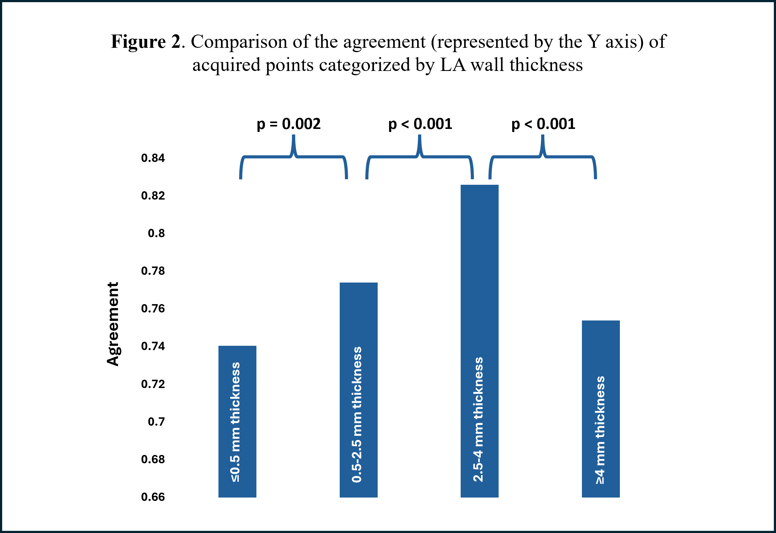
The lowest agreement was observed in the LA roof, while the highest was seen in the inferoseptal and anterior walls. Categorizing acquired points into different ranges of LA wall thickness (among 16 pts) revealed the highest agreement at diameters between 2.5-4 mm. In contrast, the agreement was lowest for diameters ≥4 mm and ≤0.5 mm (Figure 2).
Conclusion: LGE-CMR tends to underestimate the presence of fibrosis compared to EAM; however, the fibrosis that is detected is generally accurate. CT-based thickness measurements can be used for procedure preplanning to identify areas with enhanced CMR-based fibrosis accuracy. A large-scale study is required to validate these findings, considering various wall thicknesses and establishing the fibrosis threshold.
israelheart@ortra.com מייל לשאלות