Real-time arrhythmia detection and termination in a human induced pluripotent stem cell-derived cardiac tissue model using AI neural network
2Cardiology, Rambam Medical Center, Israel
Introduction
The study of cardiac arrhythmias has been hampered by the lack of in vitro human cardiac tissue models as well as by the inability to perform real-time high-resolution perturbations of the system. To tackle this challenge, we combined the use of human-induced pluripotent stem cells (hiPSC) derived cardiomyocytes, tissue engineering, optogenetic tools, and a novel automatic, artificial intelligence-based system which enables real time detection and termination of arrhythmogenic activity.
Methods and results
Stable hiPSC lines, expressing the light-sensitive cation channel CoChR, were differentiated into cardiomyocytes, seeded as large-scale hiPSC-derived cardiac cell-sheets (hiPSC-CCSs), loaded with a voltage-sensitive dye, and optically mapped. Arrhythmias were induced either spontaneously, using electrical or light-induced stimulation, with and without drug applications. Our dataset includes both healthy and disease-specific hiPSCs (demonstrating the long and short QT syndromes). This resulted in various reentrant arrhythmias (spiral waves) characterized by a different number of rotors’ cores, rotation rate, and wavelength [Fig.1a]. The neural network model was trained using 90 arrhythmia and 90 normal recordings, all were filtered and augmented [Fig.1b]. Validation accuracy after training was 92% [Fig.1c]. Python software was developed to enable optical recording of the tissue, pacing, real-time arrhythmia detection, and optogenetic termination (“defibrillation”). Real-time arrhythmia detection and termination took less then 0.5sec in all cases [Fig.1d].
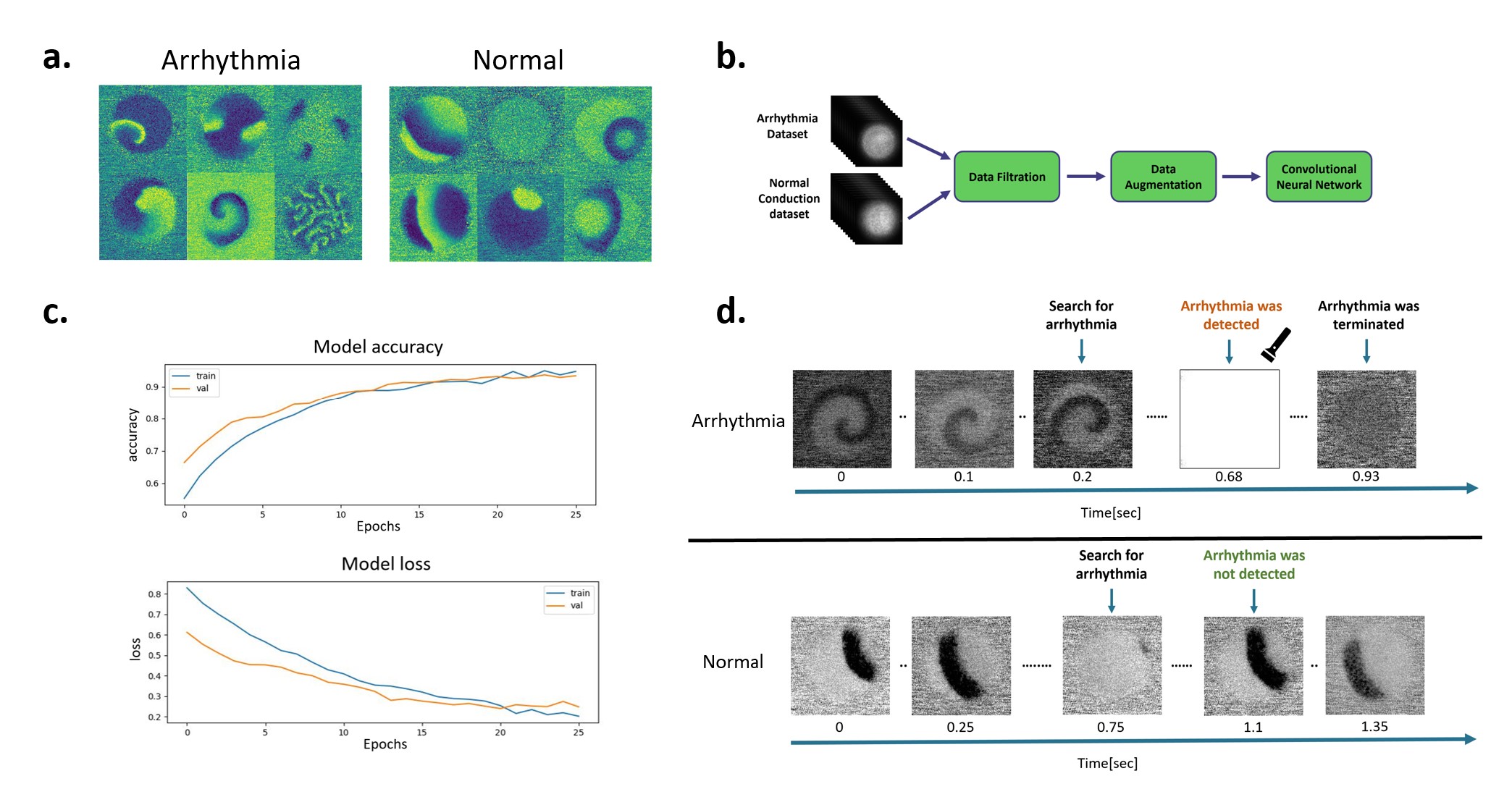
Figure 1: (a) Examples of arrhythmia and normal datasets. (b) Model workflow. (c) Model learning curves. (d) Example of real-time optogenetic arrhythmia detection and termination.
Conclusion
We developed a real-time, AI-based arrhythmia detection and termination platform using healthy and diseased hiPSC-CCSs. This innovative approach can bring a unique value to arrhythmia mechanistic studies, high throughput drug screening and disease modelling. Furthermore, it facilitates the exploration of various methods for arrhythmic activity termination and may pave the way for AI-driven solutions for arrhythmia detection and treatment in clinical setting.
israelheart@ortra.com מייל לשאלות