Advanced 3D bioprinting for cardiac regenerative medicine, disease modeling, and drug testing
2Cardiology Department, Rambam Health Care Campus, Israel
The mechanistic investigation and development of new therapies for cardiac disorders, the leading cause of worldwide morbidity and mortality, have been hampered by the lack of clinically relevant human cardiac tissue models in-vitro, specifically those reflecting the accurate 3D anatomy and cell density of the human heart as well as the patient’s specific structural and functional abnormalities. In this project, we aim to tackle these hurdles by generating anatomically relevant, patient-specific 3D tissue models of the human heart chambers.
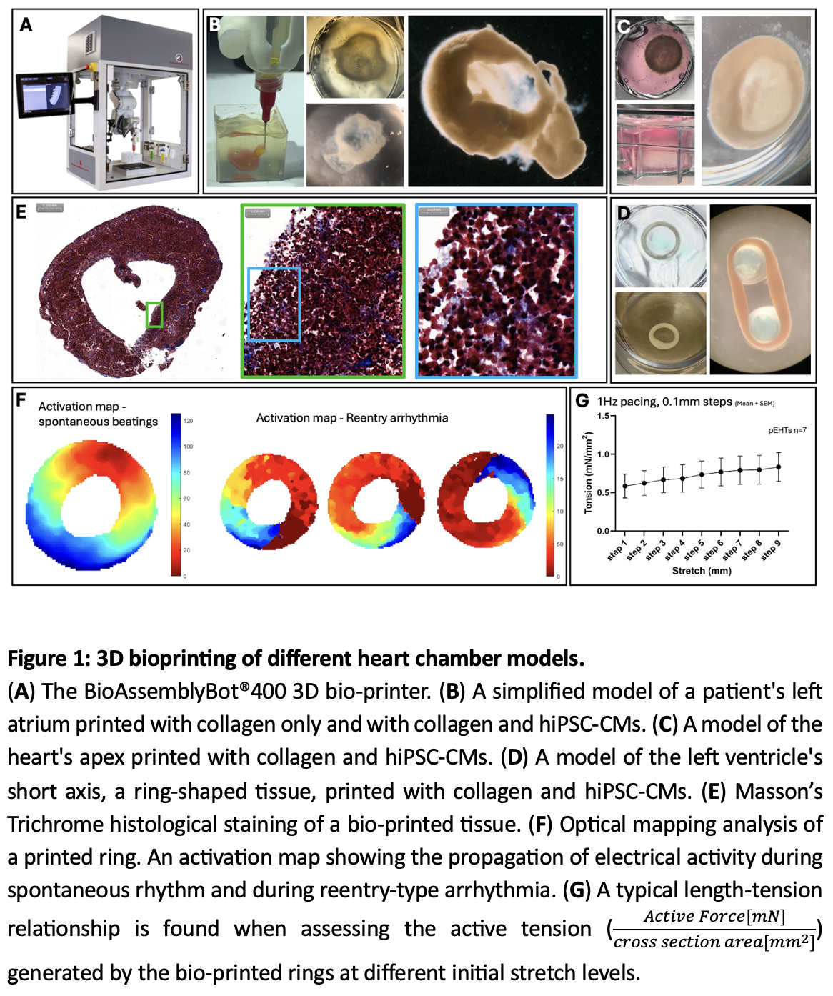
Our project combines the methods of human induced pluripotent stem cells (hiPSCs) and state-of-the-art three-dimensional (3D) bioprinting methodologies. To successfully 3D bioprint viable tissue using the BioAssemblyBot®400, two important components need to be optimized: a support bath must be used to prevent the initial collapse of the bioprinted structure, and a bioink containing a matrix component with cells. To this end, we are using a thermo-reversible gelatin microparticle support bath that possesses shear-thinning and self-healing properties, and methacrylated type I bovine collagen that can be crosslinked by light, as the fibrillar collagen type-I is the primary component of the heart`s extracellular matrix. The cells in the bioink are hiPSC-derived cardiomyocytes (hiPSC-CMs).
We have successfully bioprinted models of the left ventricle`s short axis - a ring-shaped tissue, the heart`s apex - a hemisphere, and a simplified left atrium. Our results show that the cells within the 3D-bioprinted tissue survived and beat spontaneously and synchronously. Mechanical assessment studies of the printed rings confirmed the formation of a functional syncytium that generates contractile force according to Frank Starling`s law, which describes the heart`s length-tension relationship. Optical mapping studies demonstrated the propagation of action potentials and the ability to create a reentry-type arrhythmia. Mason`s trichrome histological staining confirms a connection between the numerous cells and the uniform distribution of collagen within the engineered tissue.
israelheart@ortra.com מייל לשאלות