Optogenetic Approaches to Study Arrhythmia Mechanisms and Treatments in a Humanized Recellularized Atrial Model
2Cardiology Department, Rambam Health Care Campus, Israel
Introduction: The study of atrial arrhythmias has been limited by the lack of anatomically-relevant human cardiac tissue models and by the inability to perform targeted, functional, perturbations of the system. To engineer light-controllable clinically-relevant atrial chambers for studying arrhythmia mechanisms and therapies.
Material and method: We combined human induced pluripotent stem cells (hiPSCs) derived atrial cells, rat heart decellularization/recellularization, and optogenetic tools (hiPSCs expressing the light-sensitive depolarizing channel CoChR and high-resolution illumination device).
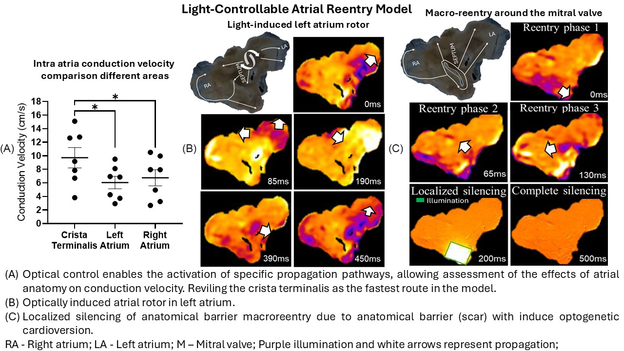
Results and discussion: We successfully generated anatomically-accurate bioengineered humanized bi-atrial tissue models. Immunostaining for chamber-specific cardiomyocyte markers, optical action-potential recordings, and the response to atrial-specific pharmacology confirmed the human atrial-specific identity of the recellularized engineered tissue. Histological examination confirmed the preservation of the macroscopic and microscopic atrial architecture. This significantly impacted the bioengineered atrial anisotropic electrophysiological properties, with conduction velocity being fastest at the crista terminalis (Fig.A). The atria could be optically controlled for optogenetic pacing, programmed stimulation, and localized electrical silencing. Atrial reentrant arrhythmias could be induced and classified into different mechanisms: macroreentrant arrhythmias due to anatomical barriers (valves), anisotropic structures (cristae-terminalis), or large scars; localized rotors acting as arrhythmia drivers; epicardial-endocardial breakthroughs; or a mixture of these mechanisms (Figs.B,C). Localized optogenetic silencing, targeting the presumed critical sites for arrhythmia perpetuation, resulted in arrhythmias’ termination; confirming the role of each mechanism (Fig.C). Finally, diffuse illumination could induce “optogenetic cardioversion” in all cases.
Conclusion: A novel, light-controllable, bioengineered humanized bi-atrial tissue model was established and could be used for atrial arrhythmia mechanistic studies and for evaluation of novel optogenetic therapeutic interventions.
israelheart@ortra.com מייל לשאלות