Brain and Spine, and Nowhere Else: An Unusual Presentation of Scedosporium Infection
Background:
Scedosporium is an opportunistic mold that may cause severe disseminated infection in immunocompromised patients, with the respiratory tract representing a common portal of entry. Central nervous system involvement is recognized, but simultaneous brain and spinal infection without an identifiable primary focus is unusual.
Case Presentation:
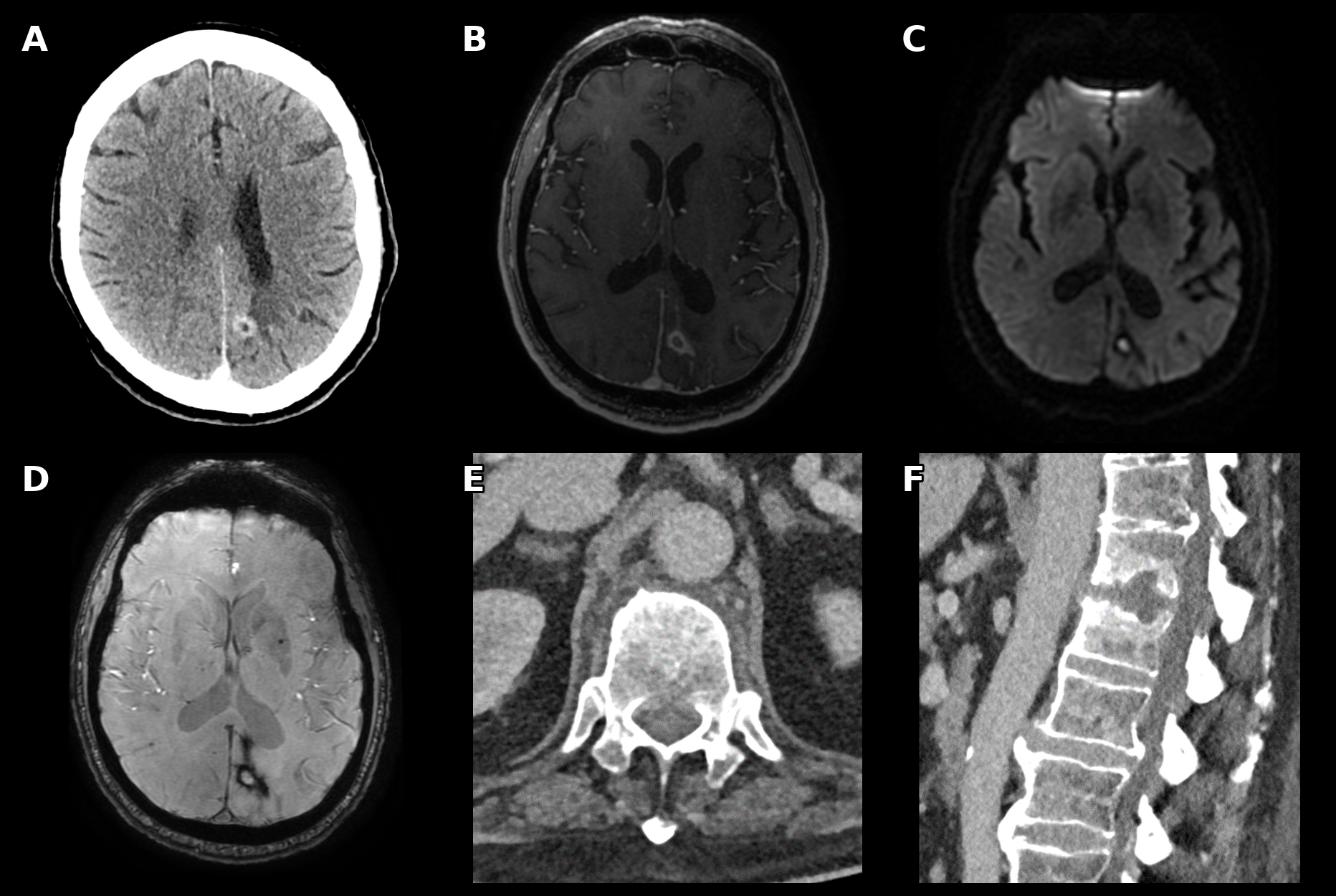
A patient with acute myeloid leukemia following hematopoietic stem-cell transplantation presented with new left lower-limb paralysis. Contrast- enhanced brain CT revealed a focal intracranial lesion (A). MRI demonstrated an irregular peripherally enhancing lesion with central diffusion restriction and peripheral susceptibility blooming, raising strong suspicion for fungal infection (B, C, D). Brain biopsy, however, was nondiagnostic.
As the location of the intracranial lesion did not explain the patient`s lower-limb weakness, spinal imaging was pursued and demonstrated D12–L1 spondylodiscitis (E, F). Chest CT and imaging of the paranasal sinuses showed no evidence of infection or potential primary source. Given the highly suspicious appearance of the brain lesion and the newly identified spondylodiscitis, the spinal lesion was biopsied. Histopathologic/microbiological evaluation confirmed Scedosporium infection.
Discussion and Conclusion:
This case illustrates an unusual presentation of Scedosporium infection involving two noncontiguous sites, the brain and spine, without an identifiable pulmonary or sinonasal source. It also highlights the importance of pursuing a second site of disease when initial tissue sampling is nondiagnostic. In immunocompromised patients, characteristic imaging findings may provide a crucial clue to invasive fungal infection and guide further diagnostic sampling.