
A Complex Pediatric Renal Abscess Mimicking a Neoplastic Process: A Diagnostic and Imaging Challenge
Background
Renal abscess is a rare cause of pediatric retroperitoneal masses that can mimic malignancy, representing a form of renal pseudotumor capable of closely simulating a tumor on imaging.
Case Presentation
A previously healthy 10-year-old girl presented with a one-month history of fever, abdominal pain, and 4 kg weight loss. Inflammatory markers were elevated with thrombocytosis, while urinalysis and urine culture were unremarkable and sterile, respectively.
Imaging Findings and Diagnosis
Ultrasound revealed a complex, multiloculated 6.0×5.3×6.2 cm mass involving the left renal upper pole and posterior pancreatic tail, without a clear separating plane; computed tomography showed an infiltrative pattern with stranding and retroperitoneal lymphadenopathy, raising concern for malignancy.
Ultrasound-guided drainage yielded purulent material, confirming abscess, and blood cultures grew Staphylococcus aureus despite sterile urine, favoring hematogenous seeding over an ascending source. Following intravenous antibiotics, the patient improved clinically, with progressive regression on serial ultrasound and magnetic resonance imaging (MRI) and near-complete resolution at one month.
Discussion and Conclusion
This case illustrates a hematogenously seeded staphylococcal renal abscess mimicking a neoplastic retroperitoneal mass, with unusual extension into the pancreatic tail. Recognizing inflammatory imaging characteristics alongside clinical signs of infection, utilizing image-guided drainage, and performing serial imaging are essential to establish an accurate diagnosis and prevent unnecessary surgical resection or biopsy.
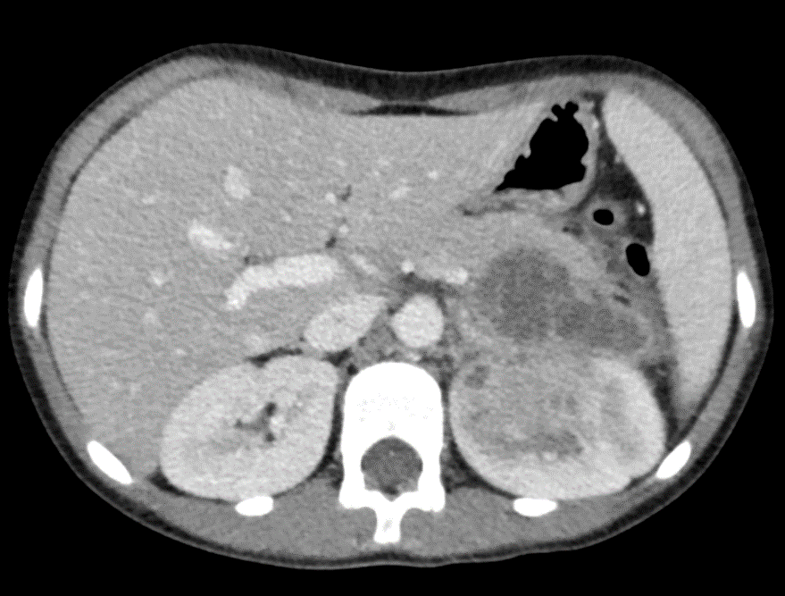
image description: Abdominal CT with IV contrast in the axial plane: multilocular, infiltrative 5.1*6.7*5.1 cm mass involving the upper pole of the left kidney and the posterior aspect of the pancreatic tail with no clear separation plane.