
Advanced 3D Segmentation-Assisted Planning for CT-Guided Cryoablation of Osteoid Osteoma
Background
3D segmentation may improve the precision and safety of osteoid osteoma cryoablation by enabling patient-specific nidus localization, trajectory planning, probe selection, and assessment of adjacent critical structures. We evaluated this workflow in anatomically challenging lesions.
Methods
Preprocedural CT-based segmentation delineated the nidus, bone, nerves, vessels, joints, and skin. Virtual needle trajectories and skin entry points were planned for direct, stable access while avoiding critical structures. Virtual ice-ball dimensions were compared with nidus coverage and predicted tissue risk. Cortical thickness and lesion dimensions were used to optimize access. The plan guided cryoprobe selection, positioning, and protective measures; when nerve injury was anticipated, an alternative drilling strategy was used (Fig.1).
Results
Eleven patients (age range, 7–26 years; 8 male, 3 female) underwent 3D planning between 2022 and 2026. Ten underwent cryoablation, involving lesions in the tibia, femur, toe, calcaneus, and fibula. Lesion size, cortical thickness, and location informed individualized probe and access planning. In a toe lesion, the ice ball reached the skin, prompting heated-saline skin protection. In a proximal fibular lesion, proximity to the common fibular nerve led to drilling instead of cryoablation (Fig 2). No major complications were documented. Postprocedural follow-up was recorded for 8/11 cryoablation patients, and no repeated cryoablation was documented.
Conclusion
Segmentation-assisted 3D planning was feasible and provided actionable information for cryoprobe selection, trajectory planning, and protection of adjacent structures. It may improve safety in complex osteoid osteoma locations, although larger studies with standardized clinical and imaging follow-up are needed.
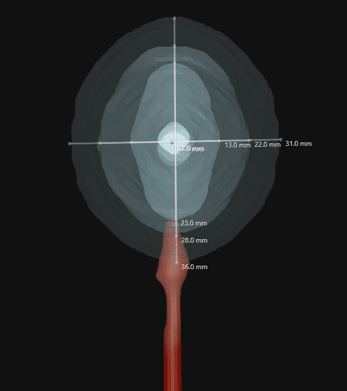
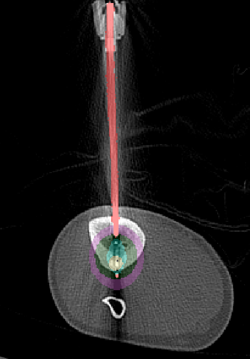
Figure 1. 3D segmentation showing the lesion, planned cryoprobe trajectory, predicted ice-ball coverage, and adjacent critical structures.
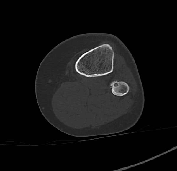
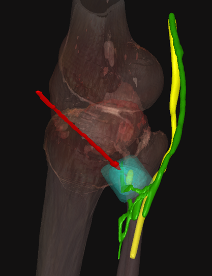
Figure 2. Axial CT shows an osteoid osteoma in the proximal fibula. 3D planning demonstrates predicted ice-ball proximity to the common fibular and tibial nerves, indicating risk of nerve injury and foot drop and prompting drilling instead of cryoablation.