
From AI-Based Segmentation to Clinical Biomarkers: An Interactive Framework for MRI Quantification of Residual Breast Tissue after Mastectomy and Silicone Implant Reconstruction
2Breast Cancer Radiation Therapy Unit, Sheba Medical Center, Tel-Hashomer, Israel
3Gray Faculty of Medical & Health Sciences, Tel Aviv University, Israel
4Meirav Center, Sheba Medical Center, Tel-Hashomer, Israel
Background: Post mastectomy monitoring requires precise quantification of residual breast tissue (RBT) thickness, which can potentially serve as an imaging biomarker for oncological surveillance and cancer recurrence. However, translating automated 3D deep learning segmentations into clinical metrics is often confounded by voxel noise, isolated tissue fragments, and boundary gaps near the chest wall or implant cavities. The objective is to develop an interactive, MRI-based framework for visualization and automated quantification of post mastectomy RBT.
Methods: Building on AI based RBT segmentations from the BRILLIANT study (utilizing an Attention U-Net architecture), we developed a three-phase pipeline: segmentation refinement, automated thickness and volume extraction, and deployment as an interactive multi-planar GUI with automated regional reporting for whole breast, RBT, and implants. Predictions undergo automated morphological filtering, inner/outer contour extraction with a convex hull fallback for missing chest walls, and 3D voxel to voxel Euclidean distance mapping. The system features click to measure distance tracking, live 3D thickness heatmaps, and automated batch processing that exports . Reports containing per-breast statistics, 3 point of interest metrics, and side specific volume analysis.
Results: The prototype successfully extracts localized thickness profiles, contour tracks, and multi component tissue volumes with sub-second interaction latency. All metrics automatically log to external templates. The pipeline generates automated thickness heatmaps over bilateral coronal slices, visualizing residual tissue distribution around reconstructive implants.
Conclusions: The framework establishes a clinically traceable pipeline that transforms binary AI segmentations into standardized geometric biomarkers. Future work will focus on multi-expert validation and evaluating the framework’s efficacy in detecting localized RBT thickness anomalies as early indicators of recurrence.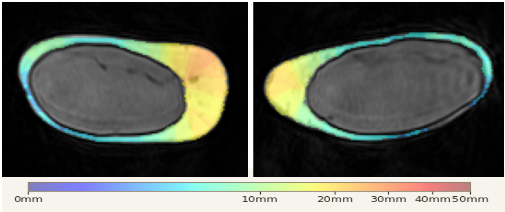
figure 1: RBT thickness heatmap (0-50+mm) with corresponding distance histogram, enabling quantitative visualization of RBT thickness distribution across the breast.