When Pathology, Imaging, and Clinical Findings Disagree: A Diagnostic Challenge
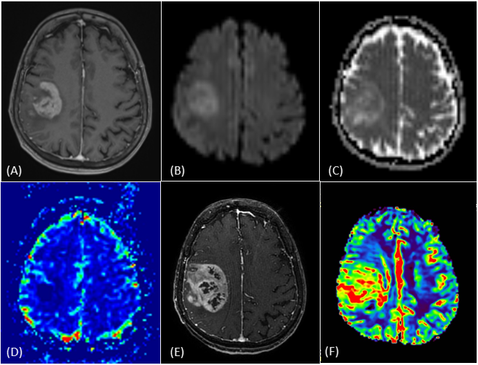
A 74-year-old patient presented with general deterioration over one week. Noncontrast CT (not shown) demonstrated an iso- to hyperdense lesion in the right frontoparietal region with surrounding edema. Initial MRI revealed an enhancing right frontoparietal lesion (A) with marked diffusion restriction on DWI and corresponding low ADC values (B, C). Perfusion imaging showed no elevation of relative cerebral blood volume (rCBV) (D).
Biopsy demonstrated inflammatory cells and material considered possibly compatible with pus, leading the pathologist to raise the possibility of infection. At that stage, these findings were considered unlikely to explain the lesion.
Following the initial MRI and biopsy, the patient deteriorated rapidly and developed a subfebrile fever. Repeat CT (not shown) demonstrated interval enlargement of the lesion within 10 days, with new areas of necrosis. Together with the biopsy findings, this raised the possibility of an infectious process superimposed on an underlying tumor and prompted repeat MRI.
The second contrast-enhanced MRI demonstrated irregular peripheral enhancement with internal areas of necrosis (E) and, in contrast to the initial examination, markedly elevated rCBV on perfusion imaging (F). These findings favored glioblastoma, and the patient underwent a second operation. Intraoperative frozen section confirmed glioblastoma, allowing extensive resection to be performed.
This case illustrates how findings suggestive of infection, together with initially negative perfusion imaging, may obscure the diagnosis of glioblastoma. When the pathological, imaging, and clinical findings are discordant, rapid evolution should prompt early repeat imaging and reconsideration of tissue sampling.