
Deep Learning for Tumor Localization in Residual Breast Tissue after Mastectomy and Reconstruction
2Breast Radiation Unit, Sheba Medical Center, Tel-HaShomer, Israel
3Gray Faculty of Medical & Health Sciences, Tel Aviv University, Israel
Background: Detecting breast cancer occurring in residual breast tissue (RBT) after mastectomy and reconstruction, whether residual or newly arising, is clinically important but challenging on dynamic contrast-enhanced magnetic resonance imaging (DCE-MRI): anatomy may be altered and enhancement is usually subtle. Automated tools for this task remain scarce. We aimed to develop an artificial intelligence (AI)-based model for localizing breast tumors in RBT under extreme data scarcity.
Methods: In this retrospective proof-of-concept study, a You Only Look Once (YOLO) detector was trained by transfer learning: pre-training on a large public breast MRI dataset, then adaptation to an imbalanced institutional cohort of 11 histologically confirmed residual tumors and 350 negative examinations. Detection was performed on maximum-intensity projections (MIP) of the subtraction series, with multiphase input and anatomy-preserving augmentation. Leave-one-out cross-validation maximized learning from the limited positive cases. Sensitivity was assessed per patient and specificity per examination.
Results: Residual tumor was detected in 7 of 11 patients (sensitivity 63.6%, 95% CI 35.4-84.8%). Mean specificity across folds was 83.4% (318 negative examinations per fold). The model consistently highlighted the tumor-site region, including cases with only partial spatial overlap (Figure 1). False-positive detections were most commonly associated with enhancing vessels and fibroglandular tissue (Figure 2).
Conclusions: An AI-based detector trained with a data-efficient strategy can localize breast cancer in RBT on post-mastectomy MRI despite extremely limited annotated data. Confidence intervals are wide given the few positive cases multicenter validation is required. With further development it may support radiologists as a second reader.
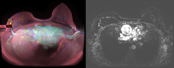
Figure 1. True positive: MIP (left) and matching subtraction slice (right). Green = pathology-confirmed tumor, red = model prediction with confidence score.
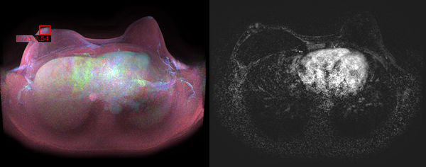
Figure 2. False positive: an enhancing focus flagged by the model with no corresponding tumor. MIP (left), matching subtraction slice (right).