Purpose: To evaluate pancreatic perfusion assessed by contrast-enhanced ultrasound in Cystic fibrosis (CF) patients with known exocrine pancreatic function.
Materials and Methods: Contrast-enhanced ultrasound was performed in CF patients (n=32) and healthy controls (n=30). Exocrine pancreatic function was assessed by secretin-stimulated endoscopic short test and/ or faecal Elastase. Perfusion data was analyzed on stored DICOM-files using DCE-US software (http://www.isibrno.cz/perfusion/) and a dedicated perfusion model. Mean transit-time (MTT), blood flow (BF) and blood-volume (BV) was calculated. Exclusions due to image quality and image analysis in the CF group were made without knowledge of pancreatic function.
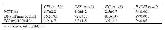
Results: 22 CF patients and 18 controls were analyzed. 10 CF patients and 12 controls were excluded due to poor image quality. The CF patients were defined as pancreas sufficient through fecal elastase >200µg/g or duodenal bicarbonate >80mmol/L. Subjects were divided as follows: CF, pancreatic insufficient (CFI, n=10), CF pancreatic sufficient (CFS, n=12) and healthy controls (HC, n=18). Results are displayed in the table (mean±SD).
Conclusion: The pancreatic insufficient CF patients had significantly longer MTT (p<0.001), lower BF (p<0.001) and lower BV (p<0.05) compared to healthy controls and pancreatic sufficient CF patients. Contrast-enhanced ultrasound can non-invasively differentiate between healthy pancreatic tissue and exocrine insufficient pancreatic tissue due to cystic fibrosis.