Colloidal particles are widely used in pharmaceutics, flow cytometric assays, biophysics, liquid toners and electronic inks, photonics and soft condensed matter. In many of these applications it is desired to know accurate particle properties such as 3D position, orientation, size, refractive index, shape and electrical charge, properties of coatings on these particles or statistical properties of many particles. We demonstrate a method based on Fourier-Bessel (FB) image moments. In this method particle images obtained by classical white light, fluorescence, confocal microscopy or any other imaging technique are decomposed into a set of 2-dimensional orthogonal FB functions with their weights, the FB coefficients. Since each image can then be represented by a point in the multidimensional space of FB-coefficients the mathematical processing becomes much easier compared to analyzing the original images. Many images of a particle at different -positions with respect to the plane of focus result in a point cloud, a ‘fingerprint’, that accurately reflects is size, refractive index. Figure 1 illustrates the basic classification of spherical colloids (PMMA and PS with different sizes) depending on their size and refractive index. An accuracy of 1% in size and 0.2% in refractive index is achieved from a single image, comparable to holographic microscopy techniques [1]. A second illustration of the method is the measurement of the retardation force in nonpolar liquids [2].
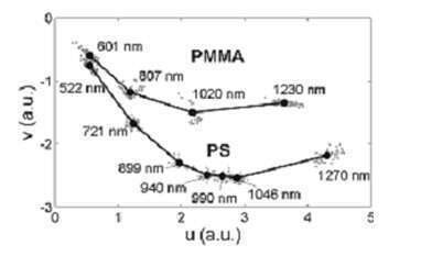
Fig. 1 Classification of individual colloidal particles depending on their refractive index and size.
[1] S-H. Lee et al., Optics express 15, 18275 (2007).
[2 ] F. Strubbe, F. Beunis, T. Brans, M. Karvar, W. Woestenborghs, K. Neyts. PRX, 021001 (2013).
filip.strubbe@elis.ugent.be