Ultrasensitive Label-Free Biosensor Based on Photon Crystal Surface Waves: a Tool to Study Dynamics of Receptor-Ligand Interactions with Living Bacteria and Cells
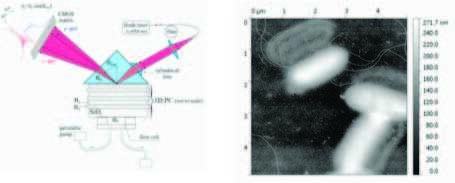
Recently, we presented a label-free biosensor based on registration of photonic crystal surface waves and discussed its first applications to study not only such “standard” systems as rabbit and mouse IgG and anti-rabbit and anti-mouse IgG protein pairs [1] but also earlier unexplored kinetics of Laminin Binding Protein (LBP) – flaviviral surface protein E interaction, which enabled (in combination with Single Molecule Dynamic Force Spectroscopy studies) unambiguously demonstrate force induced globule-coil transition of LBP and its complexes; the process which is of an uttermost importance for the viral – cell membrane fusion [2]. Angular interrogation of the optical surface wave resonance is used to detect changes in the thickness of an adsorbed layer, while an additional detection of the critical angle of total internal refection provides independent data on the liquid refraction index, see Fig. 1. Besides segregation of surface and volume effects, the exploitation of Photonic Crystal supporting long propagated surface waves enables to achieve mass sensitivity at the level of 0.3 pg/mm2 and refraction index (RI) sensitivity at the level of 10-7 RIU/Hz1/2.

Figure 1. Left: Schematic of the biosensor. Right: AFM image of (mono)layer of E. coli bacteria used as a generic “immobilized receptor layer” in the sensor.
Other characteristic feature of this biosensor is large, of the order of 1 micron, surface wave penetration depth into an external media, which enables to study intermolecular interactions not only at (a few) monolayers level, but also for such large objects as viruses, bacteria and cell organells. Here we report the first steps in this direction. We elaborated a chitosan-based protocol of surface modification of the sensor chip enabling to produce sufficiently dense and homogeneous (mono)layers of live E. coli bacteria (see Fig. above), and then these layers have been exploited as a generic “immobilized receptor layer” to study kinetics of adsorption of different ligands onto their (i.e. bacteria’s) surface.
- Konopsky, V. N. et al., Sensors, 2013, 13, 2566.
- Zaitsev, B. N. et al., J. Molecular Recognition, 2014, 27, 727.
serguei.sekatski@epfl.ch
Powered by Eventact EMS