Transparent Silica Nanopillars for SERS and LSPR Sensing Achieved by High Throughput Lithography-Free Reactive Ion Etching
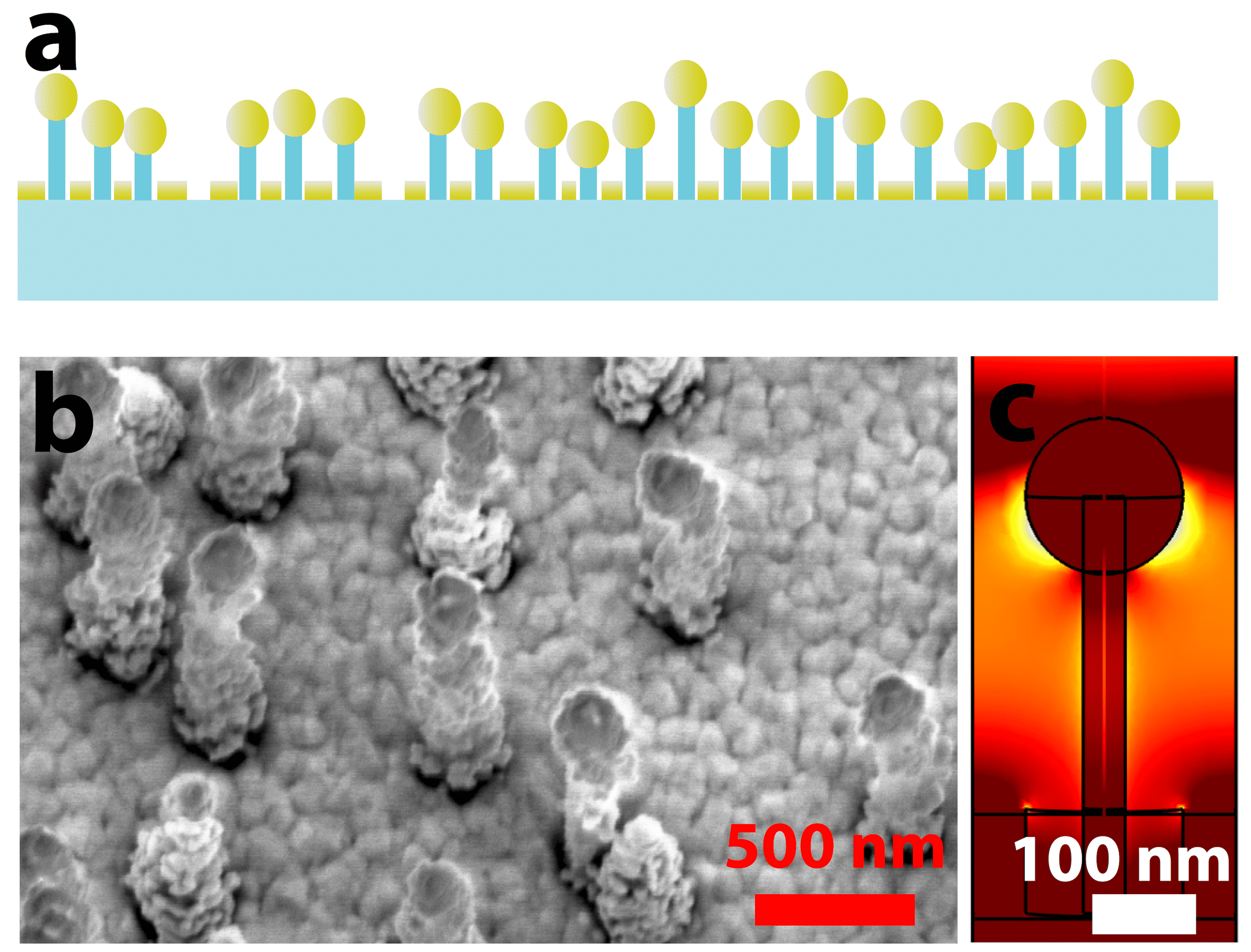
Transparent nanopillar arrays are achieved with a reactive ion etch (RIE) which does not require lithographic processing i.e. a maskless process. Depositing a thin film of gold results in a conductive structure consisting of both nanocavities and nanopillars (Figure 1a and 1b). These structures support localised surface plasmon resonance (LSPR) as well as surface plasmon polariton (SPP) modes. While retaining sufficient transparency, these structures allow for analyte detection via both reflection and transmission surface enhanced Raman spectroscopy (SERS) as well as LSPR sensing.
The fabrication procedure realizes high-density wafer scale nanopillar arrays excluding expensive process steps such as lithography. Metal coated nanopillar arrays have before been achieved in silicon using a maskless RIE for SERS detection, shown in previous work1,2. Fabrication in fused silica requires optimized SF6 and O2 gas flows as well as coil and platen power of the plasma, a process adapted and modified based on work carried out by Lilienthal et al3. Transparent metal coated nanopillar arrays with underlying nanocavities in the metal film offer unique advantages as a homogeneous plasmonic resonant surface which can be utilized to measure transmitted as well as reflected spectra. The plasmonic modes generated using a 780nm laser were simulated, and shown in Figure 1c.
SERS performance was measured with 100 µM of 1,2-bis(4-pyridyl)ethene (BPE) dissolved in ethanol. A 10X objective with a 0.1mW laser with a 780nm wavelength was used for the Raman shift measurements shown in Figure 2a, showing a S/N ratio of 177 across five measurements at different locations on the wafer. The background signal from a drop of pure water was also measured, showing a low background signal.
LSPR performance was measured using the transmitted spectrum. The extinction spectrum, in Figure 2b, shows a plasmonic absorbance peak shifting when the nanopillars are moved from air to an ethanol immersion, corresponding to a spectral sensitivity of 80 nm/RIU.


- M.S. Schmidt, J. Hübner, and A Boisen., Advanced Materials, 2012 , vol. 24, OP11-OP18.
- E.M. Hicks, S. Zou, G.C. Schatz, K.G. Spears, R.P.Van Duyne, L. Gunnarsson, T. Rindzevicius, B. Kasemo, M. Käll., 2005, Nano letters , 5.6, pp. 1065-1070.
- K. Lilienthal, M. Stubenrauch, M. Fischer, and A. Schober, 2010, Journal of Micromechanics and Microengineering , 20.2, 025017.
Powered by Eventact EMS