Capturing Molecules with Plasmonic Nanotips in Microfluidic Channels by Dielectrophoresis
The local near-field enhancement of optical nanoantennas can be exploited for different plasmonic nanosensor concepts. Surface enhanced Raman spectroscopy based on nanostructured substrates allows for chemically specific sensing with strong intensity enhancement. It has been shown that the plasmon resonances of nanocones can be engineered to fit various excitation wavelengths in the visible regime [1], and that nanocone arrays provide effective Raman platforms with the hotspots at the nanocone tips raised above the substrate [2]. One challenge in the context of surface sensing consists in delivering the analyte of interest to the location of the nanostructures and selectively attaching it to their surfaces. One possible solution lies in applying dielectrophoretic forces to the polarizable molecules by using the nanoantennas as electrodes [3-6]. Here a method for the collection and concentration of molecules on arrays of metallic nanocones is presented, making use of the high electric field gradients at the nanotips. The nanocones are integrated into a microfluidic channel and used as nanoelectrodes. By applying an AC voltage, molecules from the surrounding channel region are attracted to the tips. Simulations of the dielectrophoretic forces are presented as well as experimental proof of the proposed method [7]. After attachment of the molecules, optical read-out techniques such as Raman spectroscopy could directly be performed on the plasmonic nanostructures.

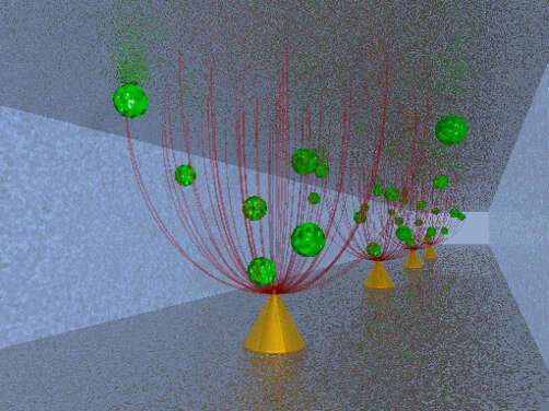
Schematic of gold nanocones in a microfluidic channel with molecules following the electric field gradient (not to scale).
[1] C. Schäfer et al., Nanoscale, 2013, 5, 7861-7866.
[2] A. Horrer et al., Small, 2013, 9, 3987-3992.
[3] I.-F. Cheng et al., Nanoscale Res. Lett., 2014, 9, 324.
[4] Y.-J. Oh and K.-H. Jeong, Lab Chip, 2014, 14, 865.
[5] S. Cherukulappurath et al., Chem. Mater., 2014, 26, 2445-2452.
[6] L. Lesser-Rojas et al., Nano Lett., 2014, 14, 2242-2250.
[7] C. Schäfer et al., Lab Chip, 2014, doi:10.1039/c4lc01018c.
monika.fleischer@uni-tuebingen.de
Powered by Eventact EMS