Background: Inflammation plays a role at all stages in the atherosclerotic process.Platelet leukocyte aggregates (PLA)present one of potential novel inflammatory markers of acute coronary syndrome (ACS). PLA includes two different cell types: platelet monocyte aggregates (PMA) and platelet neutrophil aggregates (PNA).Normal range of PLA in healthy and ACS patients is not known and utility of flow cytometry as a possible cardiovascular diagnostic technique is not established.We aim to investigate the range of PLA in normal volunteers versus patients with ACS, present Receiver Operator Characteristic (ROC) curve analysis and examine automated complete blood count (CBC) data for PLA correlation.
Methods and Results: PLA were measured blindly in 20 ACS patients and 35 healthy volunteers by flow cytometry.Ten healthy volunteer samples were taken to begin establishing normal range of PLA, examine pre-mixed cocktail stability, fluorescence controls, acquisition rate, and intra-assay repeatability.PMA measured were 26.48 +- 23.77 in ACS patients and 15.13 +- 10.42 in healthy volunteers (two independent sample T-test p=0.027).
Table 1. Statistical analysis of laboratory findings.

* Indicates one-tailed significance where assumption of increase was made previous to testing.
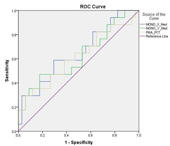
The ROC curve for the percent positive and in the PMA gate showed area under curve.
Figure 1. ROC curve analysis for PMA.

The AUC was low in all three variables, leading to a lack of significance from the null hypothesis. Abbreviations used: AUC: area under curve; MONO: monocyte; Med: Median Fluorescent Intensity; PCT: percent.
Conclusions: Patients with ACS did have higher platelet PMA compared to healthy volunteers as hypothesized. However, ROC curve analysis showed a lack of diagnostic utility, and no correlations with automated CBC were found in the variables examined. Future research goals of platelet leukocyte aggregates are discussed.
Clinical Trial Registration: NCT1797016
Key Words:
- Acute Coronary Syndrome
- Platelet Leukocyte Aggregate
- Flow Cytometry
- Cardiovascular Diagnostic Techniques

