Background: Two-dimensional strain (2DS) based on echocardiographic speckle-tracking may aid in diagnosing segmental left ventricular (LV) dysfunction, but the relative accuracy of various 2DS parameters has not been adequately established in clinical practice.
Methods: Echocardiography with 2DS imaging was performed in 54 patients (pts) hospitalized with ST-elevation myocardial infarction (MI) undergoing primary coronary angioplasty [age 56±11 years; 40 men (74%); infarct-related coronary artery (IRA) – left anterior descending: 35 pts (65%); circumflex: 7 pts (13%), right: 12 pts (22%); LV ejection fraction: 47±10%, range: 20-66%; wall motion score index: 1.67±0.26; time from admission to echocardiography: 3.3±2.4 days]. Only pts with a first MI and single-vessel coronary artery disease were included in the study. Multiple longitudinal strain parameters [peak systolic strain (pSS; at or before aortic valve closure), peak global strain (pGS; including post-systolic shortening), time-to-peak strain (TTPS)] and strain rate (SR) parameters [systolic SR (S-SR), early-diastolic SR (E-SR)] were measured by 2DS in 3 apical views (EchoPAC, GE) and compared to eyeball assessment of segmental function by a consensus of experienced echocardiographers (echocardiographic gold standard).
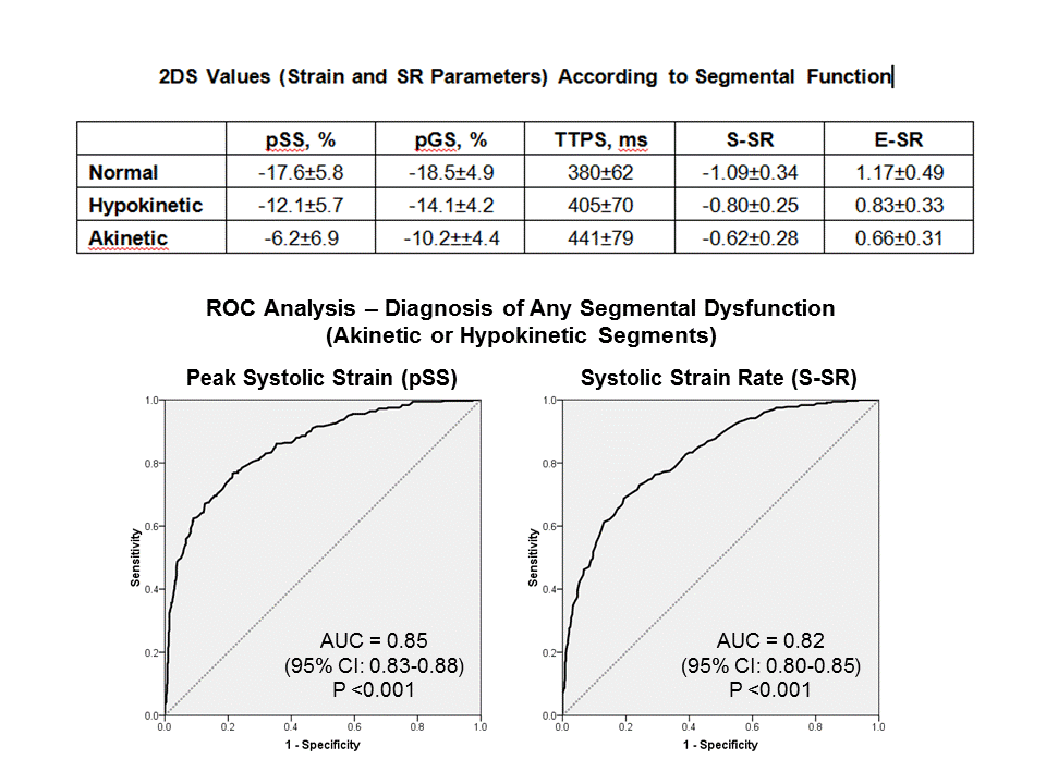
Results: 2DS measurements were feasible in 849 of 864 LV segments (98.3% feasibility), of which 490 (57.7%) segments were normal, 137 (16.1%) hypokinetic and 222 (26.1%) akinetic by visual assessment. Segmental dysfunction matched the angiographic IRA in all pts. On average, all strain and SR parameters differed significantly between normal, hypokinetic, and akinetic segments (Table), although there was considerable overlap between the normal and hypokinetic segments and between hypokinetic and akinetic segments. There was a strong correlation between various strain and SR parameters (S-SR versus pSS: r=0.77, P <0.001; E-SR versus pSS: r=-0.67, P <0.001). Using receiver operating characteristics (ROC) analysis, the area under the curve (AUC) for detecting any segmental abnormality (hypokinetic or akinetic segments; n=359 segments) by pSS was 0.85 (95% confidence interval 0.83-0.88; P <0.001) and for detecting akinetic segments was 0.88 (0.85-0.90; P<0.001) (Figures). The corresponding values of AUC for any segmental dysfunction and for akinetic segments were 0.84 and 0.86 for pGS, 0.69 and 0.72 for TTPS, 0.82 and 0.83 for SSR, and 0.70 and 0.71 for E-SR (all P values <0.001). The diagnostic accuracy was not improved by combining strain and SR parameters (pSS multiplied by S-SR; AUC of 0.85 and 0.87 for detecting any segmental dysfunction and akinetic segments).
Conclusion: Several longitudinal strain (pSS and pGS) and SR parameters (S-SR) may aid in the diagnosis of segmental LV dysfunction. There is no clear diagnostic advantage of any of these parameters over the others and no incremental value for combining strain and SR parameters.