")
A NANOSTRUCTURAL STUDY OF BLOOD SYSTEMS USING CRYOGENIC ELECTRON MICROSCOPY
2Pediatric Hematology Unit, Emek Medical Center in Afula, Afula, Israel
Blood is composed of plasma and three types of blood cells: erythrocytes, lymphocytes, and thrombocytes. The main function of the blood is to deliver cells and molecules, especially oxygen and nutrients, to different parts of the body, and remove the waste products of cell metabolism. It is also involved in maintaining homeostasis, hormone signaling, immune system functioning, and clotting. Blood is therefore the most complex and essential fluid in the body. Genetic mutations, systemic bodily disorders, or environmental factors can cause blood cell dysfunction, leading to severe diseases and, in some cases, death. Successful solutions to these problems require deep understanding of blood functioning.
The main research tools of this study are cryogenic scanning electron microscopy (cryo-SEM) combined with additional hematological research techniques. Cryo-SEM allows direct imaging of biological systems without modifying their structures. The technique can be used to study the morphology of different subpopulations that may coexist in a sample, and to understand physiological processes by imaging the system at varying stages. By using cryo-SEM, we can determine the properties of membranes as well as the internal structure of cells. We use a modern high-pressure freezing technique to prevent formation of ice crystals that may destroy fine nanostructural details. Thick (up to 200 μm) biological specimens may be prepared very close to their native state without adding cryoprotectants that might alter the cell morphology and/or viability.
The present study uses cryo-SEM to analyze the morphology of human blood cells. We initially focused on healthy cell morphology. We show the characteristic structural features, such as unique surface-connected canalicular system of thrombocytes, and membranal folds of the leucocytes. Than we have investigated morphological changes in blood cells in several diseases, including sickle-cell anemia and beta-thalassemia. Moreover, we studied the morphological changes of healthy and sick blood cells under conditions such as hypoxia and activation.
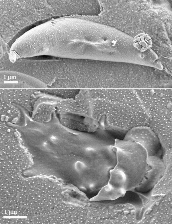
The pictures below show cryo-SEM micrographs of the RBC sample collected from a patient with sickle cell disease (HbS/β-thalassemia). The sample was incubated overnight with 15% (w/v) sodium metabisulfite to mimic the condition of hypoxia. Top: sickled cell; Bottom: acanthocyte with partially removed cell membrane. The samples were prepared using high pressure freezing. The imaging was performed on Zeiss Ultra-plus HR-SEM using ET and InLens detectors with 1:1 mixed signals. The specimens were maintained at -145 °C, and imaged without coating at an acceleration voltage of 1.2 kV, and under low-dose imaging conditions.