")
ENGINEERING THE POINT-SPREAD FUNCTION OF CONFOCAL MICROSCOPY
High-resolution subcellular imaging in patients is one of the main challenges faced by modern optical technologies, and while optical microscopy would be ideal for such task, most in-vivo microscopy techniques often suffer from subpar image quality and slow imaging rates. Today, most of the microscopy techniques used for clinical diagnosis rely on reflectance confocal microscopy (RCM), which can offer high-resolution subsurface imaging without using fluorescence labelling. Our research group studies various approaches for clinical diagnosis using RCM, and has recently demonstrated an innovative technique for non-invasive blood testing termed spectrally encoded flow cytometry (SEFC).
Here we address one of the main challenges of our SEFC system, and of RCM in general; its characteristic speckle noise and inherent interference patterns that often prevent efficient differentiation between different cell types. In order to reduce speckle noise, we suggest a new approach for engineering the point-spread function (PSF) of RCM, and demonstrate the improvement both numerically and experimentally.
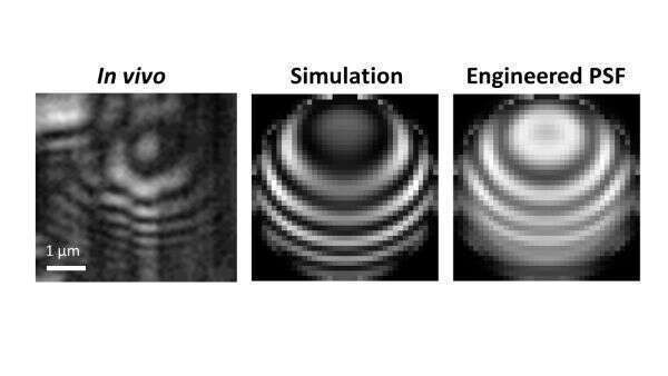
Using a dedicated Matlab code for three-dimensional simulation of the optical fields under a high numerical aperture objective lens, create a model of a red blood cell and simulate its confocal appearance in the reflectance confocal microscope. By matching the simulated image to the true SEFC images (figure 1) we are able to reconstruct the actual shape and size of the red blood cell. By applying our PSF engineering method we demonstrate that the contrast of the interference patterns formed in the red cell image can be significantly reduced, allowing smoother images that are easier to interpret and analyze.
The results of this study could improve the clinical value of SEFC, allowing improved image quality diagnosis accuracy, and may also assist other applications that require high-resolution noninvasive microscopy for effective clinical diagnosis.