Beyond the Herniated Disc: Lumbar Epidural Venous Thrombosis as a Radiologic and Diagnostic mimicker
Background: Lumbar epidural venous thrombosis is a rare cause of lumbar radiculopathy that may closely mimic intervertebral disc herniation both clinically and radiologically. Owing to its rarity and nonspecific presentation, diagnosis is often challenging and management strategies remain poorly defined.
Case Description: A 52-year-old man presented with 3–4 weeks of progressive low back pain radiating to the left lower extremity, accompanied by paresthesia and significant motor weakness (2/5 strength). D-dimer was elevated at 673 ng/mL (cutoff 500 ng/mL), with a normal complete blood count and normal coagulation screen. Lumbar magnetic resonance imaging revealed a thrombosed epidural vein at L4–L5 compressing adjacent neural structures. Ultrasonography showed no deep vein thrombosis and abdominal CT angiography showed no inferior vena cava thrombus. No clinical risk factor for thrombosis was identified. Therapeutic anticoagulation with enoxaparin 80 mg twice daily was initiated, and an evaluation for an acquired or inherited hypercoagulable state was recommended.
Conclusion: Lumbar epidural venous thrombosis is an uncommon but important differential diagnosis in patients presenting with lumbar radiculopathy and atypical epidural lesions on MRI. Early recognition may prevent misdiagnosis and unnecessary surgical intervention. Evaluation for systemic venous thrombosis and underlying hypercoagulable disorders should be considered, as identification of a predisposing cause may influence long-term management and prognosis.
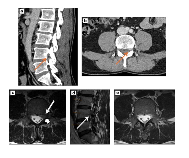
Figure 1: Lumbar spine computed tomography and magnetic resonance imaging (MRI) of a 52-year-old man with acute left-sided radiculopathy.
(A+D) Sagittal CT and T2-weighted MR image reveal severe stenosis of the left L4-5 neuroforamina caused by the extradural fullness.
(B+C) Axial CT and T2-weighted MR image at L4 demonstrates extradural fullness behind the L4 vertebral body (long arrow), along the epidural venous plexus, pressing on the L4 nerve root (short arrow image C).
(E) Follow-up axial T2-weighted MR image 6 weeks after anticoagulation shows complete resolution of the extradural fullness.